Fraud Blocker Skip to main content

Cortex Collection (page 6)

The cortex, a fascinating part of the brain, is responsible for numerous functions that are essential to our daily lives

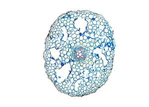
Background imageCortex Collection: Horsetail stem, light micrograph

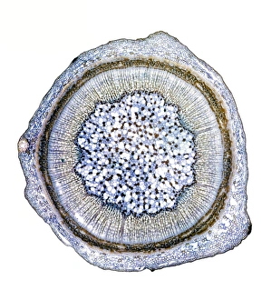
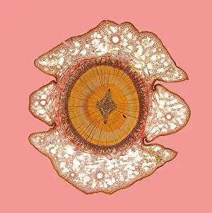
Horsetail stem, light micrograph. Transverse section through the stem of the common horsetail (Equisetum arvensis). The stem consists of an outer epidermis and an outer cortex of sclerenchyma cells

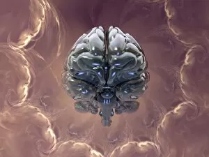
Background imageCortex Collection: Creation of the human brain, artwork

Creation of the human brain, artwork
Creation of the human brain. Conceptual computer artwork of a human brain amid swirling clouds of stardust. This could represent the creation and evolution of the modern human brain

Background imageCortex Collection: Butchers broom stem, light micrograph

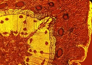
Butchers broom stem, light micrograph
Butchers broom stem. Polarised light micrograph of a section through a butchers broom stem (Ruscus aculeatis) showing many vascular bundles (dark clusters) within the plant cortex (orange)

Background imageCortex Collection: Kidney bean stem, light micrograph

Kidney bean stem, light micrograph
Kidney bean stem. Light micrograph of a section through the stem of a kidney bean (Phaseolus vulgaris) plant. The outer layer is the cuticle (brown), with a cortex of parenchyma (yellow) beneath it

Background imageCortex Collection: Lilac stem, light micrograph

Lilac stem, light micrograph
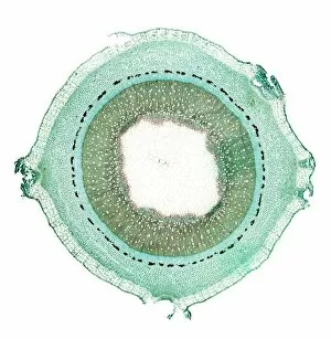
Lilac stem. Light micrograph of a transverse section through the young woody stem of a lilac (Fraxinus excelsior) tree. The thick epidermis (solid green) is being sloughed (pushed/broken)

Background imageCortex Collection: Broad bean root, light micrograph

Broad bean root, light micrograph
Broad bean root. Light micrograph of a transverse section through the young root of a broad bean (Vicia faba) plant. The outer epidermis is covered in root hairs

Background imageCortex Collection: Kidney bean root, light micrograph

Kidney bean root, light micrograph
Kidney bean root. Light micrograph of a section through the root of a kidney bean (Phaseolus vulgaris) plant. At centre is the pith (blue)

Background imageCortex Collection: Common broom stem, light micrograph

Common broom stem, light micrograph
Common broom stem. Light micrograph of a transverse section through the stem of a common broom (Salicornia europaea) plant

Background imageCortex Collection: Ash stem, light micrograph

Ash stem, light micrograph
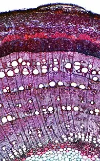
Ash stem. Light micrograph of a transverse section through the woody stem of an ash (Fraxinus excelsior) tree showing four growth rings (concentric circles)

Background imageCortex Collection: Oleander stem, light micrograph

Oleander stem, light micrograph
Oleander stem. Light micrograph of a cross-section through the stem of an oleander (Nerium oleander) tree. The outer layer is a thick cuticle (yellow) and then cork cambium (blue-green)

Background imageCortex Collection: Marsh samphire stem, light micrograph

Marsh samphire stem, light micrograph
Marsh samphire stem. Light micrograph of a transverse section through the stem of a marsh samphire, (Salicornia europaea) plant. This is a succulent, xerophytic, halophyte plant

Background imageCortex Collection: Ginkgo leaf stalk, light micrograph

Ginkgo leaf stalk, light micrograph
Ginkgo leaf stalk. Light micrograph of a transverse section through the leaf stalk (petiole) of a ginkgo tree (Ginkgo biloba)

Background imageCortex Collection: Blackcurrant plant stem, light micrograph

Blackcurrant plant stem, light micrograph
Blackcurrant plant stem. Light micrograph of a cross-section through the stem of a blackcurrant (Ribes nigrum) plant. The outer cortex (blue and pale brown) is made of cork cambium tissue

Background imageCortex Collection: Spikenard stem, light micrograph

Spikenard stem, light micrograph
Spikenard stem. Light micrograph of a cross-section through the stem of a Spikenard (Aralia sp.) tree. The dark red cells just beneath the surface bark are collenchyma cells

Background imageCortex Collection: Buttercup root, light micrograph

Buttercup root, light micrograph
Buttercup root. Light micrograph of a transverse section through the root of a buttercup (Ranunculus sp.) plant. At centre is the vascular bundle, comprising xylem (red) and phloem (blue) tissues

Background imageCortex Collection: Rose stem, light micrograph

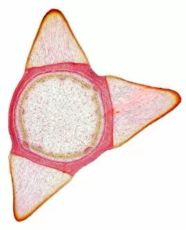
Rose stem, light micrograph
Rose stem. Light micrograph of a cross-section through the stem of a rose (Rosa sp.). The three triangular protrusions are thorns, which protect the plant from being eaten by animals

Background imageCortex Collection: Elder tree stem, light micrograph

Elder tree stem, light micrograph
Elder tree stem. Light micrograph of a transverse section through the very young woody stem of an elder (Sambuca nigra) tree

Background imageCortex Collection: Dog rose stem, light micrograph

Dog rose stem, light micrograph
Dog rose stem. Polarised light micrograph of a cross-section through the stem of a dog rose (Rosa canina) plant. The outer cortex (black and red) is made of cork cambium tissue

Background imageCortex Collection: Willow stem, light micrograph

Willow stem, light micrograph
Willow stem. Light micrograph of a cross-section through the stem of a willow (Salix sp.) tree. At centre is the pith (white), surrounded by woody xylem tissue (pink)

Background imageCortex Collection: Subconsciousness, conceptual image

Subconsciousness, conceptual image. Computer artwork of an abstract diagram above the coloured cortex of a human brain, representing the thought processes that occur in the mind

Background imageCortex Collection: Maple stem, light micrograph

Maple stem, light micrograph
Maple stem. Polarised light micrograph of a cross-section through the stem of a field maple (Acer campestre). This shows the thick layer of cork (phellem)

Background imageCortex Collection: Dutchmans pipe plant stem

Dutchmans pipe plant stem. Light micrograph of a section through the stem of a dutchmans pipe plant (Aristolochia sipho). The inner blue circle is a layer of sclerenchyma fibres

Background imageCortex Collection: Scots pine tree stem, light micrograph

Scots pine tree stem, light micrograph
Scots pine tree stem. Polarised light micrograph of a cross-section through the stem of a Scots pine tree (Pinus sylvestris), showing resin canals

Background imageCortex Collection: Stem of the nootka cypress

Stem of the nootka cypress. Light micrograph of a cross-section of the stem of the conifer nootka cypress (Chamaecyparis nootkatensis)

Background imageCortex Collection: Human brain, artwork

Human brain, artwork
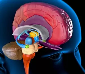
Human brain anatomy. Computer artwork of a persons head showing the brain with the right hemisphere removed. The following regions can be seen: outer cortex (red) somatosensory cortex (lighter)

Background imageCortex Collection: Human brain anatomy, artwork

Human brain anatomy, artwork
Human brain anatomy. Computer artwork of a persons head showing the brain with the right hemisphere removed. The brainstem can be seen (bottom left)

Background imageCortex Collection: Waterweed stem, light micrograph

Waterweed stem, light micrograph
Waterweed stem. Polarised light micrograph of a transverse section through a stem of the aquatic western waterweed (Elodea nuttallii) plant

Background imageCortex Collection: Mistletoe stem, light micrograph

Mistletoe stem, light micrograph
Mistletoe stem. Polarised light micrograph of a transverse section through the stem of a mistletoe (Viscum album) plant. The epidermis (outer layer)

Background imageCortex Collection: Mares tail stem, light micrograph

Mares tail stem, light micrograph
Mares tail stem. Polarised light micrograph of a transverse section through a stem of the aquatic mares tail (Hippuris vulgaris) plant. All aquatic plants (hydrophytes) have a similar stem structure

Background imageCortex Collection: Clematis stem, light micrograph

Clematis stem, light micrograph
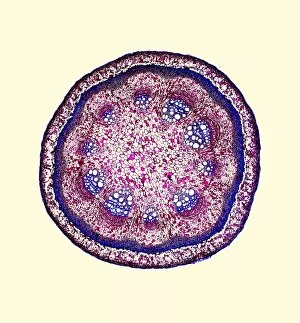
Clematis stem. Light micrograph of a transverse section through the stem of a clematis (Clematis flammula) plant. At the centre of the stem is a large area of pith, consisting of parenchyma cells

Background imageCortex Collection: Liana stem, light micrograph

Liana stem, light micrograph
Liana stem. Light micrograph of a transverse section through the stem of a liana (Aristolochia tormentosa), or woody vine. At the centre of the stem is the pith, consisting of parenchyma cells

Background imageCortex Collection: Kidney, longitudinal section

Kidney, longitudinal section
Kidney. Light micrograph of a longitudinal section through a kidney, showing the outer region (cortex, round the edges) and the inner region (medulla, centre)

Background imageCortex Collection: Brain activity, artwork

Brain activity, artwork
Brain activity. Computer artwork showing activity (red) in the frontal (left) and temporal (centre) lobes of a human brain. The front of the brain is at left


All Professionally Made to Order for Quick Shipping

The cortex, a fascinating part of the brain, is responsible for numerous functions that are essential to our daily lives. One remarkable aspect of the the motor homunculus, a representation of how different parts of our body are controlled by specific areas in the brain. This intricate map showcases the complexity and precision with which movements are coordinated. Purkinje nerve cells in the cerebellum play a crucial role in maintaining balance and coordination. These unique cells have an elaborate branching structure that allows them to transmit information efficiently within this important region of the brain. Examining a vascular bundle under scanning electron microscopy reveals its intricate network, highlighting how blood vessels supply vital nutrients and oxygen to sustain healthy brain tissue. Artwork depicting basal ganglia captures their significance as they contribute to various functions such as movement regulation and decision-making processes. Their interconnectedness emphasizes their influence on overall brain function. Understanding the complex system that ensures proper blood supply to brain tissue is crucial. The delicate balance between oxygen delivery and demand plays a critical role in maintaining optimal cognitive function. A light micrograph showcasing a lime tree stem provides insight into plant anatomy but also serves as a reminder of nature's intricate design parallels with human biology. Similar Purkinje nerve cells can be found within the cerebellum structure when observed under light microscopy. Their distinct appearance further emphasizes their importance in coordinating movements smoothly and precisely. Phantom pain after amputation remains an enigma; artwork capturing this phenomenon highlights its impact on individuals who experience sensations from missing limbs—an intriguing area where neuroscience continues to seek answers. Exploring water fern rhizome under light microscopy offers glimpses into its internal structures, reminding us that biological intricacies extend beyond humans alone—a testament to nature's diversity and complexity. A pine tree stem captured through light microscopy unveils its cellular composition—underscoring similarities between plant tissues and those found within our own bodies—a reflection of shared evolutionary history across species boundaries.