Fraud Blocker Skip to main content

Diatoms Collection

Diatoms, the mesmerizing art of nature's microscopic algae, captivate our imagination with their intricate beauty

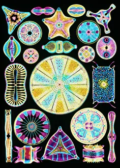
Background imageDiatoms Collection: Art of Diatom algae (from Ernst Haeckel)

Art of Diatom algae (from Ernst Haeckel)
Diatoms. Computer-enhanced engraving of assorted species of diatom. Diatoms are a group of single- celled algae

Background imageDiatoms Collection: Diatoms - marine (x25)

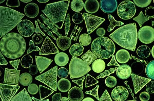
Diatoms - marine (x25)
JC-567 Diatoms - marine (x25) John Clegg Please note that prints are for personal display purposes only and may not be reproduced in any way

Background imageDiatoms Collection: Diatom - from marine plankton sample

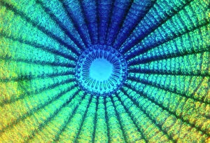
Diatom - from marine plankton sample
SPH-530 Diatom - from marine plankton sample Santa Anna, California, USA Steve Hopkin Please note that prints are for personal display purposes only and may not be reproduced in any way

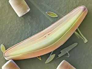
Background imageDiatoms Collection: Diatom alga, SEM

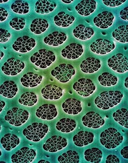
Diatom alga, SEM
Diatom. Coloured scanning electron micrograph (SEM) of the surface of the mineralised cell wall (frustule) of an unidentified diatom. This is a planktonic unicellular alga

Background imageDiatoms Collection: Picture No. 11675478

Picture No. 11675478
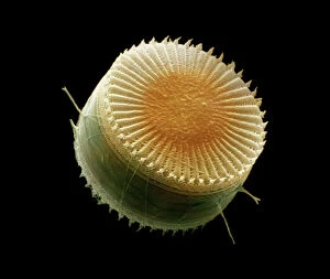
Scanning Electron Micrograph (SEM): Freshwater Diatom, Cyclotella meneghiniana. Date:

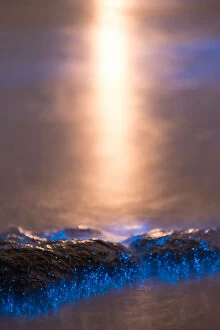
Background imageDiatoms Collection: Bioluminescence close-up

Bioluminescence close-up
Close up photograph of blue bioluminescence or Sea Sparkle caused by bioluminescent phytoplankton Noctiluca scintillans

Background imageDiatoms Collection: Diatoms, SEM

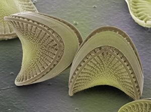
Diatoms, SEM
Diatoms. Coloured scanning electron micrograph (SEM) of diatoms (Campylodiscus sp.). These microscopic, single-celled algae possess rigid and finely-sculpted cell walls known as frustules

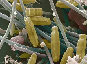
Background imageDiatoms Collection: SEM of diatoms and blue-green algae

SEM of diatoms and blue-green algae
Diatoms & blue-green algae. Coloured scanning electron micrograph (SEM) of groups of Navicula sp. diatoms (brown). Also seen in the image are strands of blue-green algae (blue)

Background imageDiatoms Collection: Fossilised diatoms, SEM

Fossilised diatoms, SEM

Background imageDiatoms Collection: Bioluminescence close-up

Bioluminescence close-up
Close up photograph of blue bioluminescence or Sea Sparkle caused by bioluminescent phytoplankton Noctiluca scintillans

Background imageDiatoms Collection: [Frustules of Diatoms], ca. 1870. Creator: Attributed to Julius Wiesner

[Frustules of Diatoms], ca. 1870. Creator: Attributed to Julius Wiesner
[Frustules of Diatoms], ca. 1870

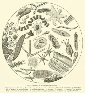
Background imageDiatoms Collection: Sediment of Water from the Thames (engraving)

Sediment of Water from the Thames (engraving)
2791441 Sediment of Water from the Thames (engraving) by English School, (19th century); Private Collection; (add.info.: Sediment of Water from the Thames)

Background imageDiatoms Collection: Types of diatoms (litho)

Types of diatoms (litho)
6013621 Types of diatoms (litho) by English School, (20th century); Private Collection; (add.info.: Types of diatoms. Illustration for The Harmsworth Encylopaedia (c 1922).); © Look and Learn

Background imageDiatoms Collection: Diatomea, 1899-1904 (colour litho)

Diatomea, 1899-1904 (colour litho)
999849 Diatomea, 1899-1904 (colour litho) by German School, (19th century); Private Collection; (add.info.: A Kunstformen der NaturA (Art Forms in NatureA, 1899-1904)

Background imageDiatoms Collection: Bioluminescence - Hobart, Tasmania

Bioluminescence - Hobart, Tasmania
Bioluminescence glowing in the waves along a beach in Tasmania with Mt. Wellington / kunanyi moonlit in background

Background imageDiatoms Collection: Marine Diatoms

Marine Diatoms
LRMC-3 Scanning Electron Micrograph (SEM): Marine Diatoms Magnification x2400 (when printed A4)

Background imageDiatoms Collection: Microscopic Entities Patterns Patterns In Nature

Microscopic Entities Patterns Patterns In Nature
Microscopic view of diatoms

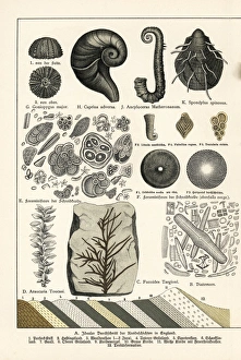
Background imageDiatoms Collection: Fossils of diatoms, foraminifera, ferns and mollusks

Fossils of diatoms, foraminifera, ferns and mollusks.. Chromolithograph from Dr. Fr. Rolles Geology and Paleontology section in Gotthilf Heinrich von Schuberts Natural History, Schreiber, Munich, 1886

Background imageDiatoms Collection: Picture No. 11014631

Picture No. 11014631
Diatom Scanning Electron Micrograph (SEM): Magnification x5275 (if print A4 size: 29.7 cm wide) - The diatoms are single celled algae

Background imageDiatoms Collection: Picture No. 11014632

Picture No. 11014632
Diatom Scanning Electron Micrograph (SEM): Magnification x 4370 (if print A4 size: 29.7 cm wide) - The diatoms are single celled algae

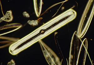
Background imageDiatoms Collection: LM of the diatom Pinnularia nobilis

LM of the diatom Pinnularia nobilis
Light micrograph of the diatom Pinnularia nobilis (centre), surrounded by other assorted diatoms. The diatoms are a distinctive group of single- celled algae containing about 10, 000 species

Background imageDiatoms Collection: Diatom - from marine plankton sample - Hong Kong harbour

Diatom - from marine plankton sample - Hong Kong harbour
SPH-552 Diatom - from marine plankton sample Hong Kong harbour Steve Hopkin Microscopic Please note that prints are for personal display purposes only and may not be reproduced in any way

Background imageDiatoms Collection: Diatom, light micrograph C014 / 4673

Diatom, light micrograph C014 / 4673
Diatom. Differential interference contrast micrograph of a Gyrosigma sp. diatom. Diatoms are a group of photosynthetic, single-celled algae containing about 10, 000 species

Background imageDiatoms Collection: Holosticha ciliate protozoan, SEM

Holosticha ciliate protozoan, SEM
Holosticha ciliate protozoan. Coloured scanning electron micrograph (SEM) of a Holosticha sp. ciliate protozoan ingesting diatoms (blue). Holosticha is a tiny single-celled aquatic organism

Background imageDiatoms Collection: Tabellaria diatoms, SEM C016 / 9599

Tabellaria diatoms, SEM C016 / 9599
Tabellaria diatoms. Coloured scanning electron micrograph (SEM) of a Tabellaria sp. diatom colony. Diatoms are a group of photosynthetic, single-celled algae containing about 10, 000 species

Background imageDiatoms Collection: Tabellaria diatoms, SEM C016 / 9600

Tabellaria diatoms, SEM C016 / 9600
Tabellaria diatoms. Coloured scanning electron micrograph (SEM) of a Tabellaria sp. diatom colony. Diatoms are a group of photosynthetic, single-celled algae containing about 10, 000 species

Background imageDiatoms Collection: Tabellaria diatoms, SEM C016 / 9601

Tabellaria diatoms, SEM C016 / 9601
Tabellaria diatoms. Coloured scanning electron micrograph (SEM) of a Tabellaria sp. diatom colony. Diatoms are a group of photosynthetic, single-celled algae containing about 10, 000 species

Background imageDiatoms Collection: Diatom frustules (SEM) C014 / 2412

Diatom frustules (SEM) C014 / 2412
Diatom frustules. Coloured scanning electron micrograph of diatom frustules (Cymbella gastroides). Diatoms are a group of photosynthetic, single-celled algae containing about 10, 000 species

Background imageDiatoms Collection: Diatom frustules (SEM) C014 / 2147

Diatom frustules (SEM) C014 / 2147
Diatom frustules. Coloured scanning electron micrograph of the internal surface of a diatom frustule of Cymbella gastroides. The tiny diatom inside the cymbella is navicula sp

Background imageDiatoms Collection: Diatoms, light micrograph C014 / 4675

Diatoms, light micrograph C014 / 4675
Diatoms. Differential interference contrast micrograph of a group of Meridion circulare diatoms. Diatoms are a group of photosynthetic, single-celled algae containing about 10, 000 species

Background imageDiatoms Collection: Diatom, light micrograph C014 / 4677

Diatom, light micrograph C014 / 4677
Diatom. Differential interference contrast micrograph of a Campylodiscus sp. diatom. Diatoms are a group of photosynthetic, single-celled algae containing about 10, 000 species

Background imageDiatoms Collection: Diatoms, light micrograph C014 / 4671

Diatoms, light micrograph C014 / 4671
Diatoms. Differential interference contrast micrograph of a group of Achnantes longipes diatoms. Diatoms are a group of photosynthetic, single-celled algae containing about 10, 000 species

Background imageDiatoms Collection: Diatom, light micrograph C014 / 4669

Diatom, light micrograph C014 / 4669
Diatom. Differential interference contrast micrograph of an Achnantes longipes diatom. Diatoms are a group of photosynthetic, single-celled algae containing about 10, 000 species

Background imageDiatoms Collection: Diatoms, light micrograph C014 / 4668

Diatoms, light micrograph C014 / 4668
Diatoms. Differential interference contrast micrograph of a group of Achnantes longipes diatoms. Diatoms are a group of photosynthetic, single-celled algae containing about 10, 000 species

Background imageDiatoms Collection: Diatoms, light micrograph C014 / 4674

Diatoms, light micrograph C014 / 4674
Diatoms. Differential interference contrast micrograph of a group of Achnantes longipes diatoms. Diatoms are a group of photosynthetic, single-celled algae containing about 10, 000 species

Background imageDiatoms Collection: Diatom, light micrograph C016 / 8606

Diatom, light micrograph C016 / 8606
Diatom. Phase-contrast light micrograph of a diatom from the Mediterranean. Diatoms are a group of photosynthetic, single-celled algae containing about 10, 000 species

Background imageDiatoms Collection: Diatoms, light micrograph C016 / 8605

Diatoms, light micrograph C016 / 8605
Diatoms, Shearing contrast light micrograph. Diatoms are a group of photosynthetic, single-celled algae containing about 10, 000 species

Background imageDiatoms Collection: Diatoms, light micrograph C016 / 8598

Diatoms, light micrograph C016 / 8598
Diatoms. Interphase contrast light micrograph of a selection of differently shaped diatoms. Diatoms are a group of photosynthetic, single-celled algae containing about 10, 000 species

Background imageDiatoms Collection: Diatoms, light micrograph C016 / 8602

Diatoms, light micrograph C016 / 8602
Diatoms. Phase-contrast light micrograph of diatoms from the Mediterranean. Diatoms are a group of photosynthetic, single-celled algae containing about 10, 000 species

Background imageDiatoms Collection: Diatoms, light micrograph C016 / 8600

Diatoms, light micrograph C016 / 8600
Diatoms. Differential interference contrast (DIC) light micrograph of diatoms from the Mediterranean. Diatoms are a group of photosynthetic, single-celled algae containing about 10, 000 species

Background imageDiatoms Collection: Diatoms, light micrograph C016 / 8585

Diatoms, light micrograph C016 / 8585
Diatoms. Interphase contrast light micrograph of two differently shaped diatoms. Diatoms are a group of photosynthetic, single-celled algae containing about 10, 000 species

Background imageDiatoms Collection: Microalgae research

Microalgae research. Biologist holding a Petri dish containing cultured diatom microalgae

Background imageDiatoms Collection: Diploneis diatom, SEM C013 / 5110

Diploneis diatom, SEM C013 / 5110
Diploneis diatom, coloured scanning electron micrograph (SEM). Diatoms are single-celled photosynthetic algae, of which there are about 100, 000 species

Background imageDiatoms Collection: LM of the colonial diatom Fragillaria sp

LM of the colonial diatom Fragillaria sp
Light micrograph of the diatom Fragillaria sp.. The filaments seen here are simple colonies of Fragillaria

Background imageDiatoms Collection: Melosira filamentous diatom alage, LM

Melosira filamentous diatom alage, LM
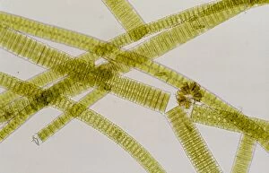
Melosira sp. diatoms. Light micrograph of filamentous strands of the freshwater diatom alga Melosira sp.. Diatoms are single-celled algae that form cell walls made of silica

Background imageDiatoms Collection: Diatom algae, Campylodiscus

Diatom algae, Campylodiscus
Diatoms. Coloured scanning electron micrograph of the diatom Campylodiscus hibernicus. The diatoms are a distinctive group of single-celled algae containing about 10, 000 species

Background imageDiatoms Collection: Colonial freshwater diatoms

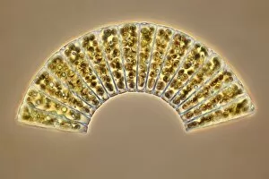
Colonial freshwater diatoms
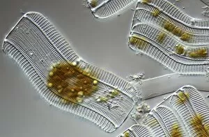
Light micrograph of assorted freshwater diatoms. The rectangular, broken filaments seen here are simple colonies

Background imageDiatoms Collection: Light micrograph of assorted diatoms

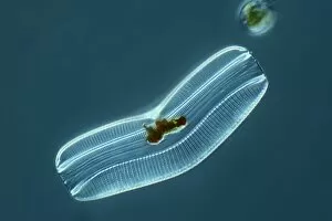
Light micrograph of assorted diatoms. The diatoms are a distinctive group of single-celled algae containing about 10, 000 species

Background imageDiatoms Collection: Diatoms - from marine plankton sample - Santa Anna - California

Diatoms - from marine plankton sample - Santa Anna - California
SPH-532 Diatoms - from marine plankton sample Santa Anna, California, USA Steve Hopkin Microscopic Please note that prints are for personal display purposes only and may not be reproduced in any way

Background imageDiatoms Collection: Diatoms - from marine plankton sample - Hong Kong harbour

Diatoms - from marine plankton sample - Hong Kong harbour
SPH-534 Diatoms - from marine plankton sample Hong Kong harbour Steve Hopkin Microscopic Please note that prints are for personal display purposes only and may not be reproduced in any way

Background imageDiatoms Collection: Marine Diatoms and Radiolarians

Marine Diatoms and Radiolarians
LRMC-17 Scanning Electron Micrograph (SEM): Marine Diatoms and Radiolaria skeletons Magnification x435 (when printed A4)

Background imageDiatoms Collection: Marine Diatom

Marine Diatom
LRDS-293 Scanning Electron Micrograph (SEM): Marine Diatom Magnification x2

Background imageDiatoms Collection: Marine Diatom

Marine Diatom
LRDS-291 Scanning Electron Micrograph (SEM): Marine Diatom Magnification x1

Background imageDiatoms Collection: Plankton - Diatom (Rhizosolenia stolterfothii) Individual diatoms unite to form

Plankton - Diatom (Rhizosolenia stolterfothii) Individual diatoms unite to form

Background imageDiatoms Collection: Diatoms, light micrograph

Diatoms, light micrograph. Diatoms are a group of photosynthetic, single-celled algae containing about 10, 000 species

Background imageDiatoms Collection: Melosira sp diatoms, light micrograph

Melosira sp diatoms, light micrograph
Diatoms. Light micrograph of Melosira monoliformis freshwater diatoms. Diatoms are a group of photosynthetic, single-celled algae containing about 10, 000 species

Background imageDiatoms Collection: Pleurosigma sp diatoms, light micrograph

Pleurosigma sp diatoms, light micrograph
Diatoms. Differential interference contrast micrograph of Pleurosigma angulatum marine diatoms. Diatoms are a group of photosynthetic, single-celled algae containing about 10, 000 species

Background imageDiatoms Collection: Fossil diatoms, light micrograph

Fossil diatoms, light micrograph. Diatoms are a group of photosynthetic, single-celled algae containing about 10, 000 species

Background imageDiatoms Collection: Pleurosigma sp diatom, light micrograph

Pleurosigma sp diatom, light micrograph
Diatom. Light micrograph of Pleurosigma angulatum marine diatoms. Diatoms are a group of photosynthetic, single-celled algae containing about 10, 000 species

Background imageDiatoms Collection: Sea cucumber plate

Sea cucumber plate. Coloured scanning electron micrograph of an ossicle (plate, orange) from a sea cucumber, surrounded by diatoms

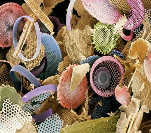
Background imageDiatoms Collection: Selection of diatoms, SEM

Selection of diatoms, SEM
Selection of diatoms, coloured scanning electron micrograph (SEM)

Background imageDiatoms Collection: Coscinodiscus diatom, SEM

Coscinodiscus diatom, SEM
Coscinodiscus diatom. Coloured scanning electron micrograph (SEM) of a Coscinodiscus diatom (red). This is a marine diatom


All products are expertly crafted, using premium materials, tailored to your specifications and promptly shipped

Diatoms, the mesmerizing art of nature's microscopic algae, captivate our imagination with their intricate beauty. Ernst Haeckel, a renowned biologist and artist, immortalized these diatom wonders through his stunning illustrations. From marine plankton samples to fossilized remains, they have left an indelible mark on our understanding of the natural world. Under the lens of a microscope at 25 times magnification, diatoms reveal themselves as delicate structures floating in the vastness of the ocean. Their symmetrical patterns and vibrant hues create a kaleidoscope that enchants scientists and artists alike. In Picture No. 11675478, we witness a close-up view captured by scanning electron microscopy (SEM). This technique allows us to explore their intricate details with unparalleled precision. The result is an awe-inspiring image that showcases the complexity hidden within these seemingly simple organisms. Fossilized diatoms provide us with glimpses into ancient ecosystems long gone. SEM imaging reveals their preserved forms, reminding us of their role as key players in Earth's history. These tiny creatures have witnessed countless changes over millions of years and continue to shape our planet today. The interplay between diatoms and blue-green algae is another captivating sight revealed by SEM imagery. In this microcosmic world, different species coexist harmoniously, creating breathtaking scenes reminiscent of abstract art. As we delve deeper into this microscopic realm through SEM technology, we uncover more secrets held by these remarkable organisms. Each new image brings forth fresh perspectives on their structure and function—reminding us that even in minuscule forms lies immense beauty waiting to be discovered. From Ernst Haeckel's artistic renderings to cutting-edge scientific techniques like SEM imaging—diatoms never cease to amaze us with their elegance and importance in shaping our planet's past and present. Through these captivating visuals, we are reminded once again that beauty can be found even in the tiniest corners of our natural world.

© Copyright Media Storehouse, All Rights Reserved 2003 - 2024
Facebook     Pinterest     100% Payment Secure     Reviews IO