Home > Science > SEM
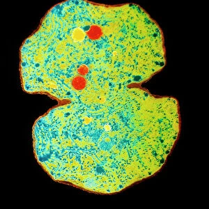
SEM of diatoms and blue-green algae

![]()
Wall Art and Photo Gifts from Science Photo Library
SEM of diatoms and blue-green algae
Diatoms & blue-green algae. Coloured scanning electron micrograph (SEM) of groups of Navicula sp. diatoms (brown). Also seen in the image are strands of blue-green algae (blue). Diatoms are single celled algae that exist in huge numbers in marine and freshwater environments. Each diatom has an intricately patterned cell wall, or frustule, made of silica and pectin. The wall is in two halves which fit neatly together. They are a major constituent of plankton. As such, they are of great importance to the food chains in their environment. Blue-green algae or cyanobacteria are linear colonies of single-celled organisms. Magnification: x610 at 6x7cm size
Science Photo Library features Science and Medical images including photos and illustrations
Media ID 9194127
© POWER AND SYRED/SCIENCE PHOTO LIBRARY
Alga Algae Algal Blue Green Algae Diatom Diatoms Phytoplankton Plankton Type
EDITORS COMMENTS
This print showcases the intricate beauty of diatoms and blue-green algae, captured through a scanning electron microscope (SEM). The image reveals groups of Navicula sp. diatoms in various shades of brown, alongside delicate strands of blue-green algae in mesmerizing hues of blue. Diatoms are single-celled algae that thrive abundantly in both marine and freshwater environments. Each diatom possesses a meticulously patterned cell wall called a frustule, composed of silica and pectin. Interestingly, this wall is divided into two halves that fit together seamlessly. As vital constituents of plankton, diatoms play a crucial role in sustaining food chains within their ecosystems. The presence of blue-green algae or cyanobacteria can also be observed within this microcosmic world. These linear colonies consist of individual single-celled organisms with distinct characteristics. With a magnification level reaching x610 at 6x7cm size, this photograph offers an extraordinary glimpse into the microscopic realm where nature's wonders unfold before our eyes. It serves as a reminder that even the tiniest organisms possess astonishing complexity and contribute significantly to the intricate tapestry of life on Earth. This stunning image from Science Photo Library invites us to appreciate the remarkable diversity and significance found within botany, specifically highlighting these fascinating types of plant-like organisms: diatoms, algal species like Navicula sp. , phytoplankton communities, as well as blue-green algae such as oscillatoria - all harmoniously coexisting
MADE IN AUSTRALIA
Safe Shipping with 30 Day Money Back Guarantee
FREE PERSONALISATION*
We are proud to offer a range of customisation features including Personalised Captions, Color Filters and Picture Zoom Tools
SECURE PAYMENTS
We happily accept a wide range of payment options so you can pay for the things you need in the way that is most convenient for you
* Options may vary by product and licensing agreement. Zoomed Pictures can be adjusted in the Cart.