
Differential Interference Collection
"Differential Interference: Unveiling the Intricacies of Cellular Warfare" In the microscopic realm, a silent battle unfolds as organisms fight for survival


All Professionally Made to Order for Quick Shipping
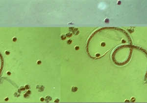
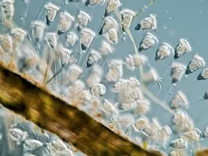
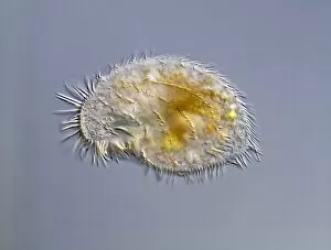
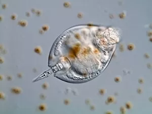
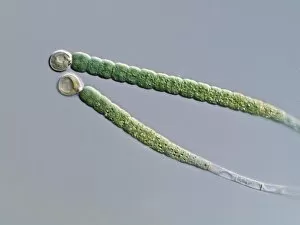
"Differential Interference: Unveiling the Intricacies of Cellular Warfare" In the microscopic realm, a silent battle unfolds as organisms fight for survival. One such example is the relentless struggle between Wuchereria bancrofti parasite and its host. Through differential interference, this parasitic worm cunningly manipulates mitosis within dividing cells, wreaking havoc on its unsuspecting victim. Under the lens of a light micrograph, Paramecium protozoa come into focus, showcasing their intricate structure and behavior. These single-celled organisms engage in an intense dance of life as they navigate their environment with grace and precision. The captivating images reveal the delicate beauty hidden within these tiny creatures. Amongst them, Vorticella protozoa captivate our attention with their unique appearance under the microscope's gaze. Their elegant stalks extend gracefully from their bodies like miniature springs ready to launch at any moment. This mesmerizing display highlights nature's remarkable diversity and adaptability. But it doesn't stop there; even more wonders await discovery in this microscopic world. A water bear emerges from obscurity, revealing its resilience against extreme conditions through its ability to survive desiccation and radiation exposure. Its tenacity serves as a reminder that life finds a way even in seemingly inhospitable environments. Pleurosigma sp diatoms add another layer of fascination to this narrative with their intricate patterns etched upon their silica shells. These beautiful structures not only serve as protection but also contribute to Earth's ecosystem by playing a crucial role in carbon cycling. Differential interference unravels these captivating stories hidden beneath our naked eyes' limitations—unlocking secrets that shed light on how organisms interact and adapt within their microscopic realms. As we delve deeper into this unseen world, we gain profound insights into the complexity of life itself—a testament to nature's ingenuity and endless surprises awaiting exploration beyond what meets our everyday sight.