
Differentiation Collection
"Unlocking the Potential: Exploring Differentiation in Stem Cells" In the vast realm of scientific research


All Professionally Made to Order for Quick Shipping
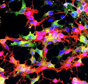
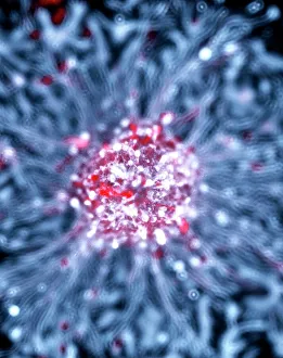
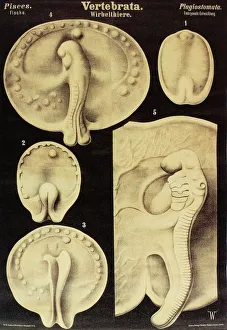
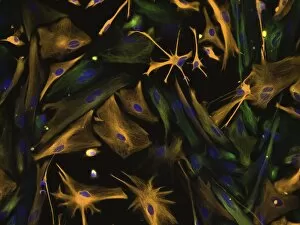
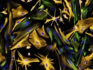
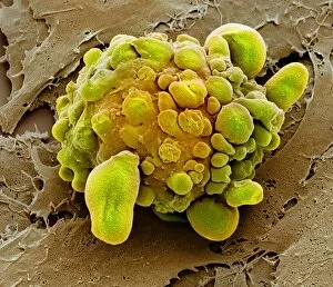
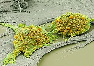
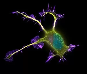
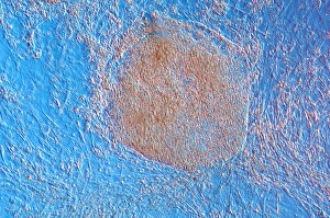
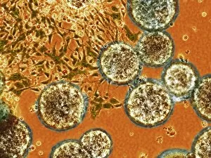
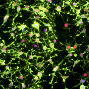
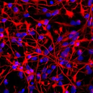
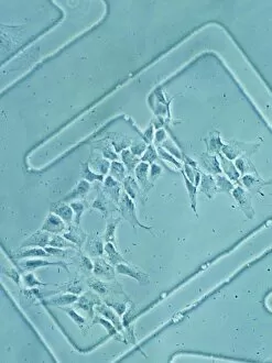
"Unlocking the Potential: Exploring Differentiation in Stem Cells" In the vast realm of scientific research, differentiation stands as a key process that holds immense promise for unlocking the potential within stem cells. Through various techniques and methodologies, scientists delve into the intricate world of cellular development, seeking to understand how these remarkable cells transform and specialize. One captivating avenue lies in glial stem cell culture, where researchers cultivate these specialized cells derived from neural tissue. Under careful observation through light micrographs, we witness their complex structures intertwining like an elaborate tapestry, offering glimpses into their unique functions within the nervous system. Similarly intriguing is neural stem cell culture – a fascinating realm where scientists nurture and study these versatile cells with boundless possibilities. As we peer through our microscope lenses at fish embryos transformed into breathtaking artwork, we marvel at nature's ability to guide cellular differentiation towards specific destinies. Mesenchymal stem cells take center stage under scanning electron microscopy (SEM), revealing their intricate features in stunning detail. These multipotent wonders hold tremendous therapeutic potential due to their ability to differentiate into various cell types such as bone or cartilage. The directed differentiation of human neural progenitor cells further captivates us as researchers skillfully coax them along specific developmental pathways towards becoming functional neurons or glial cells. The SEM images continue to amaze us as they unveil mesenchymal stem cells' diverse forms – each one representing a unique step on its journey towards specialization. We witness mesmerizing snapshots of this dynamic process; some depict vibrant clusters bursting forth with life while others showcase individual stems reaching outwards like explorers venturing into uncharted territories. Yet amidst this transformative beauty lies another facet - the delicate balance between life and death within stem cell populations. SEM images reveal stark visuals of dying stem cells – a reminder that not all paths lead to success but rather serve as stepping stones in understanding cellular fate during differentiation processes.