Fraud Blocker Skip to main content

Disease Causing Collection

"Disease-Causing Microorganisms: Unveiling the Hidden World" In a realm unseen by the naked eye

Background imageDisease Causing Collection: Salmonella bacteria, SEM

Salmonella bacteria, SEM
Salmonella bacteria, coloured scanning electron micrograph (SEM). Each rod-shaped structure is an individual bacterium. These bacteria occur mainly in human and animal intestines

Background imageDisease Causing Collection: Rift Valley fever virus, TEM

Rift Valley fever virus, TEM

Background imageDisease Causing Collection: Paramyxovirus particles, TEM

Paramyxovirus particles, TEM
Sendai virus. Coloured transmission electron micrograph (TEM) of Sendai virus particles (virions, orange). The protein coat (capsid) of one of the particles has split

Background imageDisease Causing Collection: HIV viruses, TEM

HIV viruses, TEM
HIV viruses. Coloured transmission electron micrograph (TEM) of human immunodeficiency virus (HIV) particles. These particles are docking at a T-lymphocyte (white blood cell) just before infection

Background imageDisease Causing Collection: Schistosome fluke, SEM

Schistosome fluke, SEM
Schistosome fluke. Coloured scanning electron micrograph (SEM) of a schistosome (Schistosoma sp.) fluke worm, a cause of schistosomiasis in humans

Background imageDisease Causing Collection: Schistosome flukes mating, micrograph C014 / 4867

Schistosome flukes mating, micrograph C014 / 4867
Schistosome flukes mating. Light micrograph of Schistosoma japonicum fluke worms mating. The male is the smaller animal at centre

Background imageDisease Causing Collection: Erwinia bacteria, SEM

Erwinia bacteria, SEM
Erwinia bacteria. Coloured scanning electron micrograph (SEM) of Erwinia carotovora bacteria. This gram-negative flagellated bacterium causes soft rot of plant crops such as potato

Background imageDisease Causing Collection: Naegleria fowleri protozoa, TEM

Naegleria fowleri protozoa, TEM
Naegleria fowleri protozoan. Coloured transmission electron micrograph (TEM) of a section through a Naegleria fowleri protozoan

Background imageDisease Causing Collection: Parasitic protozoan, TEM

Parasitic protozoan, TEM
Parasitic protozoan. Coloured transmission electron micrograph (TEM) of a trypanosome protozoan (Trypanosoma sp.). The nucleus (centre), cell membrane (outline)

Background imageDisease Causing Collection: Enterocytozoon sp. parasites, TEM

Enterocytozoon sp. parasites, TEM

Background imageDisease Causing Collection: Isospora sp. parasites, TEM

Isospora sp. parasites, TEM
Isospora sp. parasites. Coloured transmission electron micrograph (TEM) of Isospora sp. parasites (yellow) in a sectioned cell

Background imageDisease Causing Collection: Naegleria fowleri protozoan, TEM

Naegleria fowleri protozoan, TEM
Naegleria fowleri protozoan. Coloured transmission electron micrograph (TEM) of a section through a Naegleria fowleri protozoan

Background imageDisease Causing Collection: Mite on bee, SEM

Mite on bee, SEM
Mite on honeybee. Coloured scanning electron micrograph (SEM) of a mite (Varroa sp. upper centre) on the thorax of a honeybee (Apis mellifera)

Background imageDisease Causing Collection: Relapsing fever tick

Relapsing fever tick. Macrophotograph of a relapsing fever tick (Ornithodorus moubata) feeding on human blood. This soft-bodied tick is a parasite of humans

Background imageDisease Causing Collection: Infected bee, SEM

Infected bee, SEM
Diseased bee. Coloured scanning electron micrograph (SEM) of a honeybee (Apis mellifera) infected with mites (Varroa sp. not seen)

Background imageDisease Causing Collection: Prion protein plaque, computer artwork

Prion protein plaque, computer artwork
Prion protein plaque (right) surrounded by nerve cells (seen mainly on the left), computer artwork. Prions are infectious agents that cause a group of fatal neurodegenerative diseases

Background imageDisease Causing Collection: Adenovirus, TEM

Adenovirus, TEM

Background imageDisease Causing Collection: Dengue fever virus particles, TEM

Dengue fever virus particles, TEM

Background imageDisease Causing Collection: Dengue fever virus replication, TEM

Dengue fever virus replication, TEM

Background imageDisease Causing Collection: Malaria ookinete, TEM

Malaria ookinete, TEM
Malaria parasite. Image 5 of 10. Coloured transmission electron micrograph (TEM) of a sexual malaria ookinete in a mosquito gut wall

Background imageDisease Causing Collection: Influenza virus, TEM

Influenza virus, TEM
Influenza virus. Coloured transmission electron micrograph (TEM) of an influenza ( flu) virus particle. Influenza belongs to the orthomyxovirus group of viruses

Background imageDisease Causing Collection: West Nile viruses, TEM

West Nile viruses, TEM

Background imageDisease Causing Collection: Vaccinia virus particles, TEM

Vaccinia virus particles, TEM

Background imageDisease Causing Collection: Polio viruses, TEM

Polio viruses, TEM
Polio virus. Coloured transmission electron micrograph (TEM) of a section through a para- crystalline array of polio virus particles

Background imageDisease Causing Collection: Yellow fever virus particles, TEM

Yellow fever virus particles, TEM
Yellow fever virus particles. Coloured transmission electron micrograph (TEM) of yellow fever virus particles (white) growing in culture

Background imageDisease Causing Collection: Crimean-Congo haemorrhagic fever virus

Crimean-Congo haemorrhagic fever viruses. Coloured transmission electron micrograph (TEM) of Crimean- Congo haemorrhagic fever (CCHF) viruses (brown)

Background imageDisease Causing Collection: Bovine prion protein, molecular model

Bovine prion protein, molecular model
Bovine prion protein. Computer model showing the molecular structure of a bovine (cow) prion protein. Prions are abnormal proteins that cause a group of fatal neurodegenerative diseases including BSE

Background imageDisease Causing Collection: Legionella bacteria, SEM

Legionella bacteria, SEM
Legionella bacteria. Coloured scanning electron micrograph (SEM) of Legionella pneumophila bacteria, the cause of Legionnaires disease

Background imageDisease Causing Collection: Legionella bacteria, light micrograph

Legionella bacteria, light micrograph
Legionella bacteria. Coloured light micrograph of Legionella pneumophila bacteria, the cause of Legionnaires disease. This rod-shaped

Background imageDisease Causing Collection: Legionella bacterium, TEM

Legionella bacterium, TEM
Legionella bacteria. Coloured transmission electron micrograph of a Legionella pneumophila bacterium, the cause of Legionnaires disease

Background imageDisease Causing Collection: Mycobacterium bovis, TEM

Mycobacterium bovis, TEM
Mycobacterium bovis. Coloured transmission electron micrograph (TEM) of a group of M. bovis bacteria. It is a Gram-positive, aerobic, non- motile bacterium. M. bovis is a variant of M

Background imageDisease Causing Collection: Gas gangrene bacteria, SEM

Gas gangrene bacteria, SEM
Gas gangrene bacteria (Clostridium perfringens), coloured scanning electron micrograph (SEM). These rod-shaped bacteria are pathogenic

Background imageDisease Causing Collection: Multiresistant bacterium

Multiresistant bacterium
Multiresistant bacterim, computer artwork. Bacterium with antibiotic resistance due to genetic mutations. This bacterium is resistant to several antibiotics and is known informally as a superbug

Background imageDisease Causing Collection: Borrelia bacteria, SEM

Borrelia bacteria, SEM
Borrelia bacteria, coloured scanning electron micrograph (SEM). These bacteria (Borrelia sp.) are pathogenic, causing diseases in humans and in animals

Background imageDisease Causing Collection: Diphtheria bacteria, TEM

Diphtheria bacteria, TEM
Diphtheria bacteria. Coloured transmission electron micrograph of a section through a group of Corynebacterium diphtheriae bacteria, the causative agent of the disease diphtheria

Background imageDisease Causing Collection: Erwinia bacteria, TEM

Erwinia bacteria, TEM
Erwinia bacteria. Coloured transmission electron micrograph (TEM) of the soil bacterium Erwinia carotovora carotovora. This gram-negative flagellated bacterium causes soft rot of plant crops such as


All Professionally Made to Order for Quick Shipping

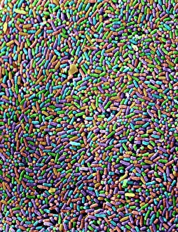
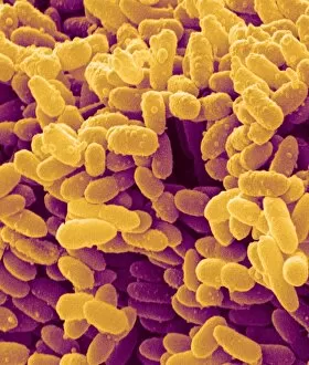
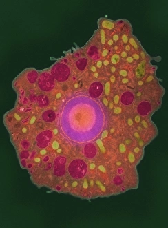
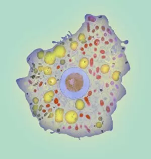
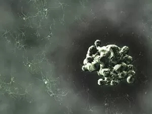
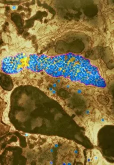
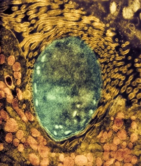
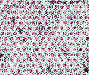
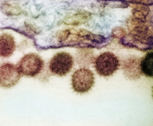
"Disease-Causing Microorganisms: Unveiling the Hidden World" In a realm unseen by the naked eye, lies a world teeming with microscopic organisms that have the power to wreak havoc on our health. Meet Salmonella bacteria, notorious for causing foodborne illnesses and gastrointestinal distress. Under the scanning electron microscope (SEM), their rod-shaped bodies appear like tiny invaders ready to strike. Moving on to Rift Valley fever virus, its presence can be detected through transmission electron microscopy (TEM). This viral culprit is responsible for outbreaks of severe fever and hemorrhagic diseases in both humans and animals. Its intricate structure reveals a complex network of proteins designed to infiltrate host cells. Enter Malaria parasites, another formidable enemy lurking within our bloodstream. TEM exposes their delicate yet deadly nature as they invade red blood cells, causing recurring bouts of high fevers and fatigue that plague millions worldwide. Paramyxovirus particles take center stage next under TEM's lens. These respiratory viruses are known for causing various ailments such as measles and mumps, showcasing an array of shapes resembling miniature spaceships poised for attack. Dengue fever virus particles follow suit in TEM imagery; these spherical entities are responsible for spreading dengue fever through mosquito bites. Their distinct spikes serve as key markers distinguishing them from other viral culprits. Adenovirus takes us deeper into the microscopic world with its hexagonal capsid structure observed via TEM. Known to cause respiratory infections and conjunctivitis among others, this virus demonstrates how even seemingly simple geometries can harbor immense danger. HIV viruses come into focus next under TEM's powerful magnification capabilities. These infamous retroviruses target immune cells relentlessly, leading to AIDS if left unchecked. Their unique shape showcases their ability to evade detection while silently compromising our immune system's defenses. Switching gears slightly from pathogens to parasites brings us Schistosome fluke seen through SEM imaging techniques—these flatworms cause the debilitating disease schistosomiasis.