Fraud Blocker Skip to main content

Disease Collection (page 100)

"Disease: A Historical Battle Against the Unseen Foe" In 1845, as the HMS Erebus and HMS Terror set sail on their ill-fated Arctic expedition

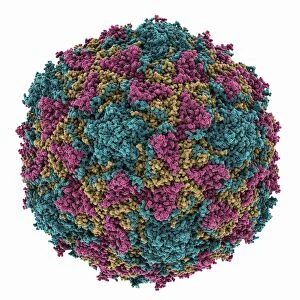
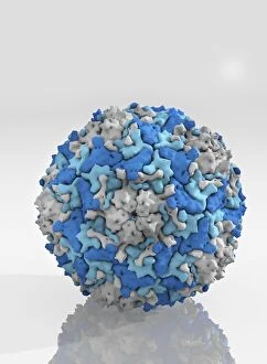
Background imageDisease Collection: Simian rotavirus capsid, molecular model

Simian rotavirus capsid, molecular model. This virus is named for its ability to infect the higher primates (simians). Rotaviruses, transmitted by faecal-oral contact

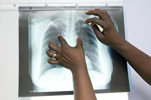
Background imageDisease Collection: Tuberculosis X-ray diagnosis

Tuberculosis X-ray diagnosis
MODEL RELEASED. Tuberculosis (TB) X-ray diagnosis. Close-up of an X-ray of a patients lungs being examined by medical personnel for signs of TB

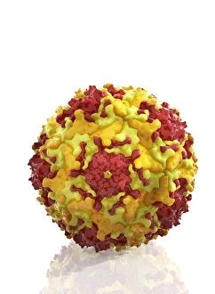
Background imageDisease Collection: Poliovirus type 3 capsid, molecular model

Poliovirus type 3 capsid, molecular model. This enterovirus causes poliomyelitis (polio) in humans, which affects the nervous system, sometimes leading to paralysis

Background imageDisease Collection: Beta-amyloid peptide molecule, artwork C014 / 2657

Beta-amyloid peptide molecule, artwork C014 / 2657
Molecular ribbon representation of the beta-amyloid peptide. The amyloid fibrillar form is the primary component of amyloid plaques found in the brains of Alzheimers disease patients

Background imageDisease Collection: Urine diabetes test C014 / 1230

Urine diabetes test C014 / 1230
MODEL RELEASED. Urine diabetes test. Woman using a urine self-test for diabetes. This test detects the levels of glucose and ketone in the urine

Background imageDisease Collection: Epstein-Barr virus protein bound to DNA C014 / 0875

Epstein-Barr virus protein bound to DNA C014 / 0875
Epstein-Barr virus protein bound to DNA. Computer model showing a molecule of Epstein-Barr nuclear antigen 1 (EBNA1) bound to a strand of DNA (deoxyribonucleic acid)

Background imageDisease Collection: Small bowel obstruction, X-ray C014 / 1819

Small bowel obstruction, X-ray C014 / 1819
Small bowel obstruction. Coloured X-ray of the abdomen of a 77 year old male patient with a small bowel obstruction and gallstones (spheres at top left). The white areas are gas-filled bowel loops

Background imageDisease Collection: HeLa cells, SEM C014 / 0369

HeLa cells, SEM C014 / 0369
HeLa cells. Coloured scanning electron micrograph (SEM) of HeLa cells (round) grown in a 3D matrix (background). HeLa cells are a continuously cultured cell line of immortal human cancer cells

Background imageDisease Collection: Rectal polyp removal, artwork

Rectal polyp removal, artwork
Rectal polyp removal. Artwork showing the use of a colonoscope to remove a polyp or cyst from the inside of the rectum. The rectum (down left) is the final part of the digestive system

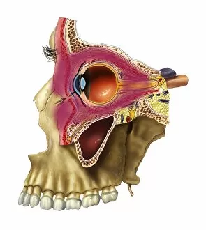
Background imageDisease Collection: Plegmon eye inflammation, artwork

Plegmon eye inflammation, artwork
Plegmon eye inflammation. Artwork of a sagittal section through a human eye and the surrounding structures, in a case of a subcutaneous (below the skin) phlegmon inflammation of an eye cavity

Background imageDisease Collection: Uterine fibroid, X-ray C014 / 4919

Uterine fibroid, X-ray C014 / 4919
Uterus tumour. Coloured X-ray of the abdomen of a woman with a fibroid (round, centre) in her uterus. A fibroid is a fibrous benign tumour originating from muscular tissue (leimyoma)

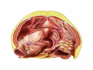
Background imageDisease Collection: Ovarian cyst in abdomen, artwork

Ovarian cyst in abdomen, artwork
Ovarian cyst in abdomen. Artwork of a large ovarian cyst (centre left) in a dissected human abdomen, seen from above. The uterus (centre) and the ovaries are part of the female reproductive system

Background imageDisease Collection: Enterovirus particle C014 / 4900

Enterovirus particle C014 / 4900
Enterovirus particle. Computer artwork of an enterovirus particle (virion), showing the structure of the capsid (outer shell)

Background imageDisease Collection: Uterine fibroid, X-ray C014 / 4918

Uterine fibroid, X-ray C014 / 4918
Uterus tumour. Coloured X-ray of the abdomen of a woman with a fibroid (orange, round) in her uterus. A fibroid is a fibrous benign tumour originating from muscular tissue (leimyoma)

Background imageDisease Collection: Enterovirus capsid proteins structure C014 / 4897

Enterovirus capsid proteins structure C014 / 4897
Enterovirus capsid proteins structure. Computer artwork showing how the four component proteins (VP1 to VP4) of an enterovirus particle (virion) interlock to form the capsid (outer shell)

Background imageDisease Collection: Patient going into oxygen chamber

Patient going into oxygen chamber
Oxygen therapy. A patient about to be loaded into a small chamber used in hyperbaric oxygen therapy (HBOT). Here a flexible chamber is being used in the treatment of a pateint with a heart condition

Background imageDisease Collection: Endometriosis, X-ray C014 / 4917

Endometriosis, X-ray C014 / 4917
Endometriosis. Coloured X-ray of the abdomen of woman with endometriosis. Endometriosis is the movement of cells from the lining of the uterus to the abdominal cavity

Background imageDisease Collection: Enterovirus capsid proteins structure C014 / 4896

Enterovirus capsid proteins structure C014 / 4896
Enterovirus capsid proteins structure. Computer artwork showing how the four component proteins (VP1 to VP4) of an enterovirus particle (virion) interlock to form the capsid (outer shell)

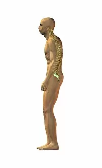
Background imageDisease Collection: Spinal disorder onset, artwork

Spinal disorder onset, artwork
Spinal disorder onset. Artwork of a man standing in a hunched posture demonstrating the onset of deteriorating spinal alignment

Background imageDisease Collection: Endometriosis, X-ray C014 / 4916

Endometriosis, X-ray C014 / 4916
Endometriosis. Coloured X-ray of the abdomen of woman with endometriosis. Endometriosis is the movement of cells from the lining of the uterus to the abdominal cavity

Background imageDisease Collection: Enterovirus particle C014 / 4898

Enterovirus particle C014 / 4898
Enterovirus particle. Computer artwork of an enterovirus particle (virion), showing the structure of the capsid (outer shell)

Background imageDisease Collection: Heart chamber wall defect, artwork

Heart chamber wall defect, artwork
Heart anatomy. Artwork of a posterior (from behind) view of a normal heart, sectioned to show the internal anatomy. The red arrow shows the normal flow of blood from the left ventricle (at left)

Background imageDisease Collection: Mouth cancer cells, SEM

Mouth cancer cells, SEM
Mouth cancer cells. Coloured scanning electron micrograph (SEM) of squamous cell carcinoma (cancer) cells from a human mouth

Background imageDisease Collection: Mouth cancer cell dividing, SEM

Mouth cancer cell dividing, SEM
Mouth cancer dividing. Coloured scanning electron micrograph (SEM) of a squamous cell carcinoma (cancer) cell from a human mouth undergoing mitosis (nuclear division)

Background imageDisease Collection: Schistosome flukes mating, micrograph C014 / 4867

Schistosome flukes mating, micrograph C014 / 4867
Schistosome flukes mating. Light micrograph of Schistosoma japonicum fluke worms mating. The male is the smaller animal at centre

Background imageDisease Collection: Narrowed artery, artwork

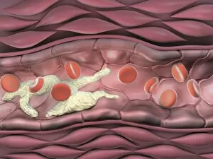
Narrowed artery, artwork
Narrowed artery. Computer artwork of a section through an artery (horizontal, centre) affected by atherosclerosis, showing red blood cells (red) and fat deposits (cream)

Background imageDisease Collection: Enterovirus particles C014 / 4899

Enterovirus particles C014 / 4899
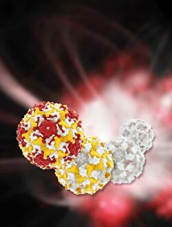
Enterovirus particles. Computer artwork of enterovirus particles (virion). Enteroviruses are a genus of non-enveloped positive-sense single-stranded RNA viruses associated with several human

Background imageDisease Collection: Wart structure, artwork

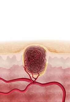
Wart structure, artwork
Wart structure. Artwork of a section through human skin, showing the subsurface blood vessels (red) supplying a wart. Warts are harmless

Background imageDisease Collection: Spinal disorder, artwork

Spinal disorder, artwork
Spinal disorder. Artwork of a man standing in a markedly hunched posture demonstrating the onset of deteriorating spinal alignment
Background imageDisease Collection: Frozen shoulder ligaments, artwork

Frozen shoulder ligaments, artwork
Frozen shoulder ligaments. Artwork of a frontal view of the bones and ligaments (blue) of a human shoulder joint, showing the condition known as a frozen shoulder (adhesive capsulitis)

Background imageDisease Collection: Infected shoulder capsule, artwork

Infected shoulder capsule, artwork
Glenohumeral capsule, infected, schematic description

Background imageDisease Collection: Mouth cancer cell, SEM

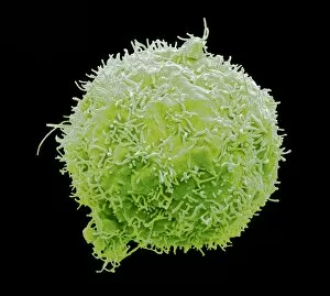
Mouth cancer cell, SEM
Mouth cancer cell. Coloured scanning electron micrograph (SEM) showing the surface of a squamous cell carcinoma (cancer) cell from a human mouth

Background imageDisease Collection: Liver cancer cell, SEM

Liver cancer cell, SEM
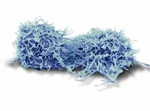
Liver cancer cell. Coloured scanning electron micrograph (SEM) of a hepatocellular carcinoma (HCC) cell, showing the numerous filopodia (hair-like) covering its surface

Background imageDisease Collection: Dividing liver cancer cell, SEM

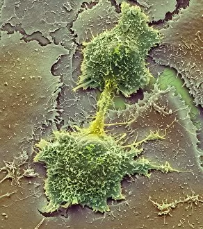
Dividing liver cancer cell, SEM
Dividing liver cancer cell. Coloured scanning electron micrograph (SEM) of a hepatocellular carcinoma (HCC) cell undergoing mitosis (nuclear division)

Background imageDisease Collection: Medical imaging in lung disease research

Medical imaging in lung disease research. Radiologist preparing a gamma camera and computed tomography (CT) scanner to produce 3D images of a patients lungs

Background imageDisease Collection: Pancreatic cancer, artwork C016 / 8735

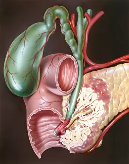
Pancreatic cancer, artwork C016 / 8735
Pancreatic cancer. Artwork of part of the digestive system, showing a carcinoma (white, lower right) of the head of the pancreas (light brown)

Background imageDisease Collection: Gastrointestinal disorders, artwork C016 / 8734

Gastrointestinal disorders, artwork C016 / 8734
Gastrointestinal disorders. Artwork of part of the digestive system, showing ulcers on the internal lining of the stomach (centre right) and the internal lining of the duodenum (centre left)
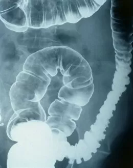
Background imageDisease Collection: Colonic spasm, X-ray

Colonic spasm, X-ray
Colonic spasm. Double contrast coloured X-ray of a spasm in the large intestine (colon). The spasm is the narrowed area at left. This could be caused by irritable bowel syndrome

Background imageDisease Collection: Stomach cancer, barium X-ray

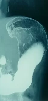
Stomach cancer, barium X-ray
Stomach cancer. Coloured frontal X-ray of the upper gastroduodenal tract of a patient with stomach cancer (grey, upper centre)

Background imageDisease Collection: Atherosclerosis, X-ray

Atherosclerosis, X-ray
Atherosclerosis. X-ray of a patient with atherosclerosis of the abdominal aorta (white, down centre). The aorta is the bodys main artery, it carries blood from the heart to the rest of the body

Background imageDisease Collection: Diseased cocoa pods, Ecuador C016 / 8421

Diseased cocoa pods, Ecuador C016 / 8421
Diseased cocoa pods. Cocoa (Theobroma cacao) fruit (pods) infected with frosty pod rot disease. This damaging disease is caused by the basidiomycete fungus Moniliophthora roreri

Background imageDisease Collection: Bowel disease in the colon C016 / 8338

Bowel disease in the colon C016 / 8338
Bowel disease in the colon. Endoscopic view of diverticular disease (diverticulosis) affecting the sigmoid colon. Diverticulosis causes sacs (diverticula)

Background imageDisease Collection: Secondary bowel cancer, endoscopic view C016 / 8337

Secondary bowel cancer, endoscopic view C016 / 8337
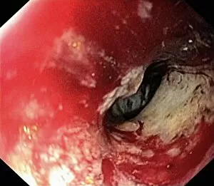
Secondary bowel cancer. Endoscopic view of the interior of a patients descending duodenum (first part of the small intestine), showing cancer that has spread (metastasised) from the pancreas

Background imageDisease Collection: Intestinal lymphoid hyperplasia C016 / 8334

Intestinal lymphoid hyperplasia C016 / 8334
Intestinal lymphoid hyperplasia. Endoscopic view of lymphoid hyperplasia in the terminal ileum - the last part of the small intestine

Background imageDisease Collection: Cold sore C014 / 4603

Cold sore C014 / 4603
Cold sore. Close-up of a cold sore beneath the nose of a 27-year-old female patient. Cold sores are caused by the Herpes simplex virus

Background imageDisease Collection: Hiatal hernia, endoscopic view C016 / 8330

Hiatal hernia, endoscopic view C016 / 8330
Hiatal hernia. Endoscopic view of a hiatal hernia (gastro-oesophageal prolapse), seen from below, with the endoscope(black) seen in an inverted position inside the stomach

Background imageDisease Collection: Hiatal hernia, endoscopic view C016 / 8329

Hiatal hernia, endoscopic view C016 / 8329
Hiatal hernia. Endoscopic view of a hiatal hernia (gastro-oesophageal prolapse), seen from above. A hernia is the protrusion of an organ, or the fascia of an organ

Background imageDisease Collection: Hyperplastic polyps in the stomach C016 / 8327

Hyperplastic polyps in the stomach C016 / 8327
Hyperplastic polyps in the stomach. Endoscopic view of small hyperplastic polyps (growths) in the gastric fundus of the stomach


All Professionally Made to Order for Quick Shipping

"Disease: A Historical Battle Against the Unseen Foe" In 1845, as the HMS Erebus and HMS Terror set sail on their ill-fated Arctic expedition, little did they know that disease would become their deadliest adversary. The Shotley Bridge General Hospital in County Durham witnessed countless patients afflicted by mysterious ailments brought back from distant lands. Captain Francis Crozier of HMS Terror valiantly led his crew through treacherous waters, but even his courage could not shield them from the invisible enemy lurking within. As depicted in a Punch cartoon, renowned scientist Faraday tried to offer his expertise to Father Thames, symbolizing society's desperate attempts to combat diseases. The battle against disease takes place at microscopic levels too. T lymphocytes engaging cancer cells under an electron microscope (SEM C001 / 1679) exemplify our ongoing fight against this relentless foe. Awareness campaigns remind us that "Coughs and Sneezes Spread Diseases, " urging us to take precautions for public health. Medical advancements have revolutionized treatments over time. Pneumothorax treatment using X-rays has saved countless lives by effectively collapsing lung cavities caused by diseases like tuberculosis. However, these triumphs cannot overshadow the tragedies faced by Captain Sir John Franklin and his crew who succumbed to disease during their Arctic exploration. Diseases have often been personified throughout history – King Cholera presiding over a court reminds us of the devastating cholera outbreaks that plagued communities worldwide. Medical illustrations depicting appendicitis or bunions captured the urgency with which doctors sought solutions for these painful conditions. Yet perhaps one of the most feared diseases is Alzheimer's, which silently erodes memories and identities. Computer artwork visualizing an Alzheimer's brain serves as a stark reminder of its impact on individuals and families alike. As we navigate through centuries battling various diseases, it becomes evident that our fight is far from over.