
Dislocation Collection
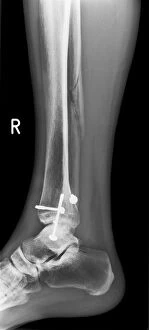
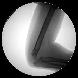
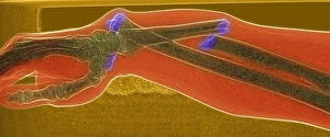
"Dislocation: A Journey of Healing and Restoration" In the realm of human existence, dislocation is an undeniable reality that can manifest in various forms


All Professionally Made to Order for Quick Shipping
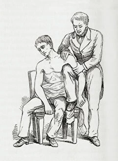
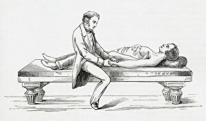
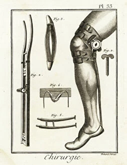
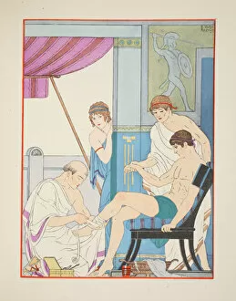
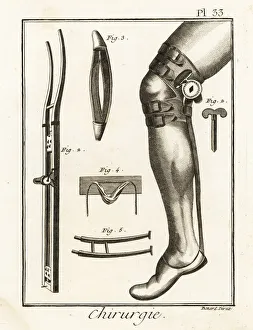
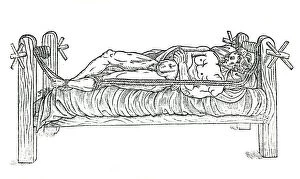
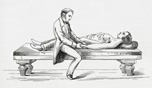
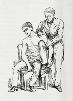
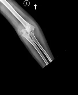
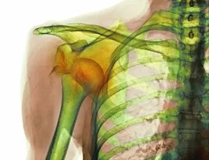
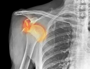
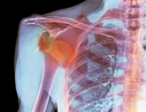
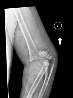
"Dislocation: A Journey of Healing and Restoration" In the realm of human existence, dislocation is an undeniable reality that can manifest in various forms. From the wounded soldier on a battlefield to the intricate illustrations from The Works of Hippocrates, it has left its mark throughout history. One such portrayal takes us back to the 18th century, where surgical knee pads and dislocation machines were employed to mend fractured bones. These archaic devices stand as testaments to humanity's relentless pursuit of finding solutions for physical ailments. The Luxation sous coracoidienne de l'épaule illustration captures a moment frozen in time - a vivid depiction of the displacement of the humerus head. Its vibrant colors serve as a reminder that even amidst pain and suffering, art can illuminate our understanding of medical conditions. Moving forward through time, we encounter apparatuses designed specifically for reducing dislocations; one dating back to 1544 showcases early attempts at rectifying this bodily misalignment. Fast forward centuries later, Sopwith Model V reveals how even coal strata can experience dislocations - highlighting that nature itself is not immune from these disruptions. But it is within ourselves that we find both resilience and innovation. The Sopwith Model III demonstrates our ability to navigate through life's challenges by addressing the dislocations within strata formations. Turning towards more specific instances, we delve into methods for reducing shoulder and elbow dislocations without assistance or with unconventional aids like heels or knees placed strategically in armpits. These techniques harken back to c. 1898 when The Household Physician published invaluable knowledge on self-care during times when professional help may be out of reach. As we conclude this journey through time, let us reflect upon an 18th-century surgical knee pad and dislocation machine alongside a page from Surgical instrument catalogue (c. 1900). They remind us that progress comes hand-in-hand with ingenuity - pushing boundaries and expanding our understanding of the human body.