
Duodenum Collection (#3)
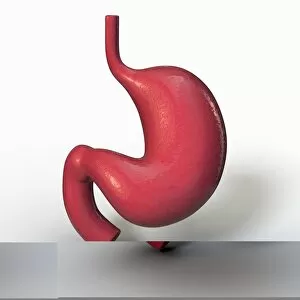
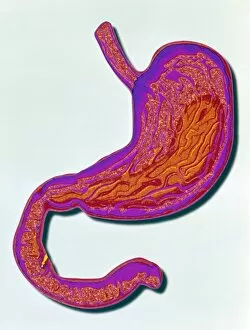
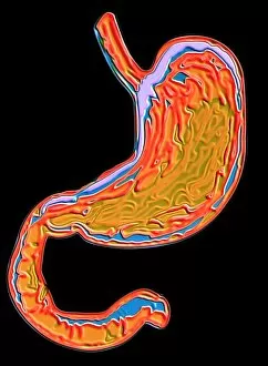
The duodenum, a vital component of the human digestive system, plays a crucial role in the gastric bypass procedure


All Professionally Made to Order for Quick Shipping
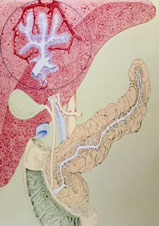
The duodenum, a vital component of the human digestive system, plays a crucial role in the gastric bypass procedure. This cross-section biomedical illustration showcases its connection to the esophagus and highlights its significance in digestion. In this captivating artwork depicting dog anatomy, we can observe the intricate placement of the duodenum within their digestive system. It serves as a testament to how animals share similar internal structures with humans. Moving on to the female body, an enlightening image reveals both her digestive and circulatory systems. The duodenum takes center stage, reminding us of its importance in breaking down food and absorbing nutrients. Meanwhile, an anatomical depiction of male respiratory organs offers insight into how they coexist alongside other internal structures like the duodenum. This comprehensive view allows us to appreciate our bodies' complexity. Similarly, an exploration of female anatomy unveils not only reproductive organs but also internal structures such as the duodenum. Understanding these intricacies helps us grasp how different systems work together harmoniously. Delving deeper into our bodies' inner workings, we encounter a mesmerizing visualization showcasing both respiratory and digestive organs side by side. The presence of the duodenum emphasizes its integration within these essential bodily functions. A vivid color lithograph provides a detailed glimpse into abdominal and thoracic internal organs where we find key players like liver, pancreas, and indeed -the ever-present duodenum. These interconnected components ensure optimal bodily function. Examining specific regions more closely brings attention to critical relationships between stomach, liver, and most importantly-our trusty companion-the duodenum. Their collaboration is fundamental for efficient digestion processes. Focusing solely on pancreatic anatomy sheds light on yet another facet involving this remarkable organ's interaction with surrounding structures including our steadfast friend-the duodenum. Finally, a 3D rendering grants us access to healthy female internal organs. This visual marvel gives prominence not only to reproductive parts but also highlights the duodenum's position within this intricate network of systems.