
Ecto Parasite Collection
"Exploring the Intricate World of Ecto Parasites: From Head Lice to Fleas and Beyond" Delving into the microscopic realm


All Professionally Made to Order for Quick Shipping
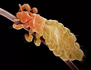
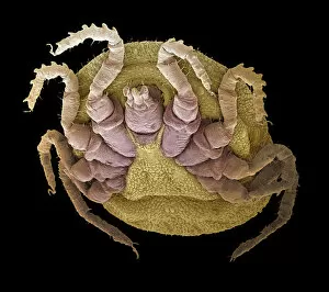
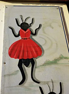
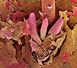
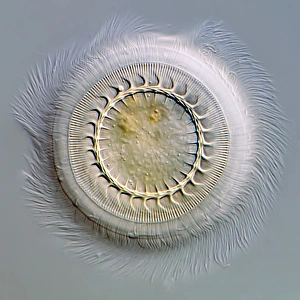
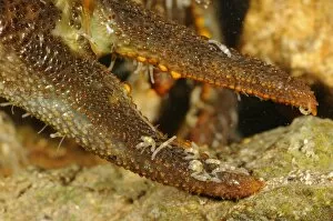
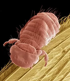
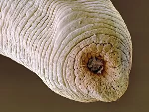
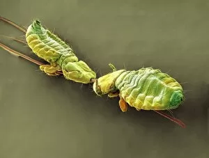
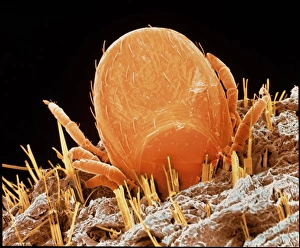
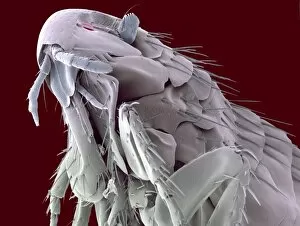
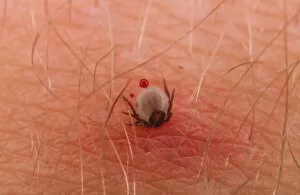
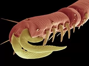
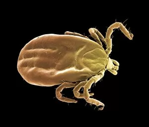
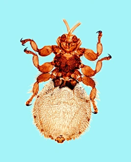
"Exploring the Intricate World of Ecto Parasites: From Head Lice to Fleas and Beyond" Delving into the microscopic realm, we uncover a captivating world teeming with ecto parasites. Through scanning electron microscopy (SEM), we witness the minuscule yet formidable head louse, its tiny claws gripping onto human hair strands like a tightrope walker defying gravity. Moving on, our attention shifts to the sheep tick, another notorious ecto parasite that attaches itself firmly onto its host's skin. Under SEM, its intricate mouthparts resemble a menacing apparatus designed for bloodsucking survival. But not all ecto parasites evoke fear; some offer intriguing spectacles. Enter the flea circus, where these agile creatures perform acrobatic feats under magnification. Their vibrant colors and delicate structures astound as they leap through miniature hoops and balance on invisible tightropes. Venturing further into this hidden world reveals unexpected wonders. Eyelash mite tails come into focus under SEM, resembling wispy appendages that navigate their way amidst eyelashes without detection by their unsuspecting hosts. Intriguingly grotesque yet medically significant is the medicinal leech – an ancient remedy for various ailments. Its segmented body and powerful suction cups are revealed in vivid detail through SEM imagery. Stepping away from SEM but no less fascinating is the light micrograph showcasing Trichodina parasite - a circular organism with cilia adorning its surface like ethereal hairs dancing in water droplets. For those seeking unconventional curiosities within this realm of parasites, look no further than the colored SEM image capturing a flea penis in all its peculiar glory – an anatomical marvel tailored for reproduction. Zooming out slightly from individual organisms brings us to fleas mating captured under SEM - an intimate moment frozen in time as nature perpetuates itself through these tiny insects' union. Shifting gears slightly but still within this captivating ecosystem, we encounter the white-clawed freshwater crayfish.