Fraud Blocker Skip to main content

Electron Micrograph Collection (page 9)

"Unlocking the Hidden World: Exploring Electron Micrographs" Delve into the microscopic realm and witness the intricate beauty of life through electron micrographs

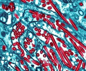
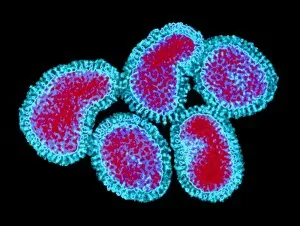
Background imageElectron Micrograph Collection: Avian flu virus, TEM

Avian flu virus, TEM
Avian flu. Coloured transmission electron micrograph (TEM) of influenza A virus particles (red). This is strain H5N1, which usually infects poultry, but began infecting humans in Hong Kong in 1997

Background imageElectron Micrograph Collection: Hepatitis A viruses, TEM

Hepatitis A viruses, TEM
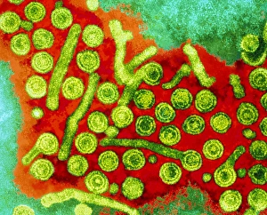
Hepatitis A viruses. Coloured transmission electron micrograph (TEM) of hepatitis A viruses, a cause of liver inflammation. Hepatitis A is transmitted through infected food or drink

Background imageElectron Micrograph Collection: Hepatitis B viruses, TEM

Hepatitis B viruses, TEM
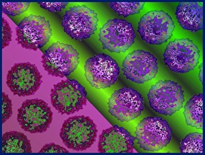
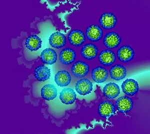
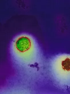
Hepatitis B viruses. Coloured transmission elect- ron micrograph (TEM) of hepatitis B viruses (large lime green circles), a cause of liver inflamm- ation

Background imageElectron Micrograph Collection: Coloured SEM of surface of an EPROM silicon chip

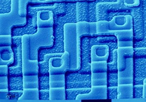
Coloured SEM of surface of an EPROM silicon chip
EPROM silicon chip. Coloured Scanning Electron Micrograph (SEM) of the etched surface of part of an EPROM silicon chip. Here

Background imageElectron Micrograph Collection: False-colour SEM connector wires on silic

False-colour SEM connector wires on silic
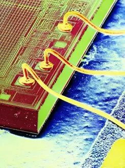
False-colour scanning electron micrograph of a TM 2716 integrated circuit, or silicon chip, show- ing 2 connecting wires bonded to terminal pads on the edge of the device

Background imageElectron Micrograph Collection: SEM of the surface of an integrated chip

SEM of the surface of an integrated chip
Scanning electron micrograph of the surface of an integrated circuit from a computers arithmetic unit, the part of the central processor responsible for arthimetical and logical operations

Background imageElectron Micrograph Collection: False-colour SEM of an integrated circuit

False-colour SEM of an integrated circuit
False-colour scanning electron micrograph of a TM 2716 integrated circuit, showing a corner of the device with surrounding connecting pin leads

Background imageElectron Micrograph Collection: Coloured SEM of a single-electron transistor

Coloured SEM of a single-electron transistor
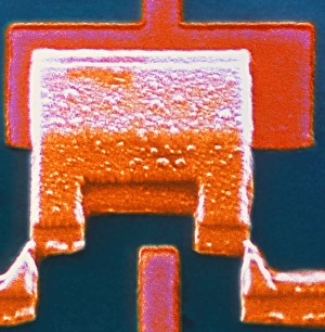
Single-electron transistor. Coloured scanning electron micrograph (SEM) of a single-electron transistor. Like the larger transistors it is designed to replace, it has three parts

Background imageElectron Micrograph Collection: Cilia in cross section

Cilia in cross section
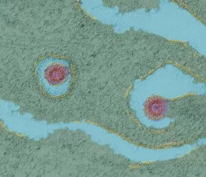
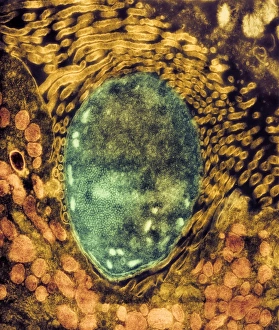
Cilia. Coloured transmission electron micrograph (TEM) of a cross section through cilia (circles), from the lining (epithelium) of the human trachea, or windpipe

Background imageElectron Micrograph Collection: Tendon fibres, TEM

Tendon fibres, TEM
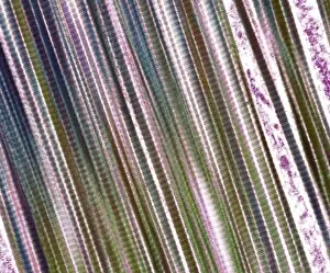
Tendon fibres, coloured transmission electron micrograph (TEM). A tendon is made up of parallel bundles of collagen fibres

Background imageElectron Micrograph Collection: Adenovirus, TEM

Adenovirus, TEM

Background imageElectron Micrograph Collection: Insect viruses, TEM

Insect viruses, TEM
Insect viruses. Coloured transmission electron micrograph (TEM) of unidentified isometric insect viruses. These viruses most commonly infect their insect hosts when they feed. Magnification unknown

Background imageElectron Micrograph Collection: Moth virus

Moth virus

Background imageElectron Micrograph Collection: Porcine endogenous retrovirus, TEM

Porcine endogenous retrovirus, TEM

Background imageElectron Micrograph Collection: Ebola virus replication, TEM

Ebola virus replication, TEM

Background imageElectron Micrograph Collection: Dengue fever virus particles, TEM

Dengue fever virus particles, TEM

Background imageElectron Micrograph Collection: Dengue fever virus replication, TEM

Dengue fever virus replication, TEM

Background imageElectron Micrograph Collection: Malaria ookinete, TEM

Malaria ookinete, TEM
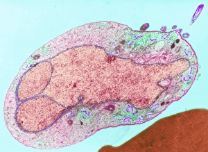
Malaria parasite. Image 5 of 10. Coloured transmission electron micrograph (TEM) of a sexual malaria ookinete in a mosquito gut wall

Background imageElectron Micrograph Collection: Malaria parasite, TEM

Malaria parasite, TEM
Malaria parasite. Image 4 of 10. Coloured transmission electron micrograph (TEM) of a sexual male malaria (Plasmodium sp.) microgametocyte in a mosquito (Anopheles sp.) gut

Background imageElectron Micrograph Collection: Cryptosporidiosis, TEM

Cryptosporidiosis, TEM

Background imageElectron Micrograph Collection: AIDS pneumonia infection, TEM

AIDS pneumonia infection, TEM
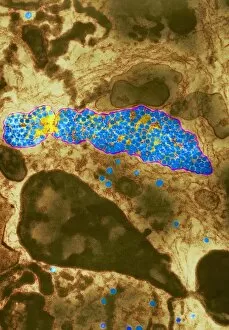
AIDS pneumonia infection. Coloured transmission electron micrograph (TEM) of a section through Pneumocystis carinii protozoa causing pneumonia. The protozoa are crescent-shaped (red/yellow)

Background imageElectron Micrograph Collection: Mosaic plant viruses, TEM

Mosaic plant viruses, TEM
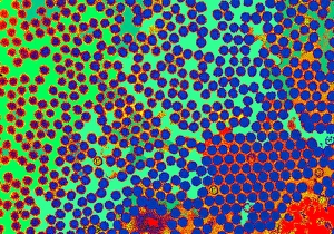
Turnip yellow mosaic viruses. Coloured transmission electron micrograph (TEM) of turnip yellow mosaic viruses (TYMV). This virus infects crucifer plants such as turnip, swede and cauliflower

Background imageElectron Micrograph Collection: Potato X virus, TEM

Potato X virus, TEM
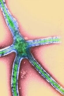
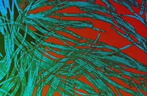
Potato X virus. Coloured transmission electron micrograph (TEM) of particles (green/blue) of the potato X virus. Potato X is a member of the potexvirus group that infects potato plants

Background imageElectron Micrograph Collection: Influenza viruses

Influenza viruses. Coloured transmission electron micrograph (TEM) of influenza (flu) viruses. The flu virus belongs to the orthomyxovirus group of viruses which have an affinity for mucus

Background imageElectron Micrograph Collection: Adenoviruses

Adenoviruses

Background imageElectron Micrograph Collection: Ebola virus, TEM

Ebola virus, TEM
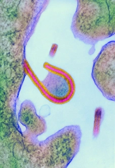
Ebola virus. Coloured transmission electron micrograph (TEM) of the extrusion (release) of an Ebola virus (red) from a host cell (green)

Background imageElectron Micrograph Collection: Hantavirus bursting from a cell

Hantavirus bursting from a cell
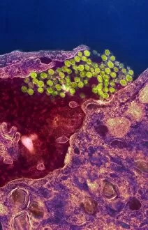
Hantavirus infection. Coloured transmission electron micrograph (TEM) of hantavirus particles (yellow/green) bursting from a human cell

Background imageElectron Micrograph Collection: Hantavirus

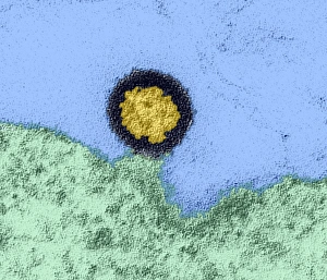
Hantavirus. Coloured transmission electron micrograph (TEM) of a hantavirus. This causes haemorrhagic fever with renal syndrome (HFRS) or Hantavirus pulmonary syndrome (HPS)

Background imageElectron Micrograph Collection: AIDS virus particle, TEM

AIDS virus particle, TEM
AIDS virus particle. Coloured transmission electron micrograph (TEM) of a section through an AIDS virus particle leaving a host cell (green, lower frame)

Background imageElectron Micrograph Collection: Polio viruses, TEM

Polio viruses, TEM
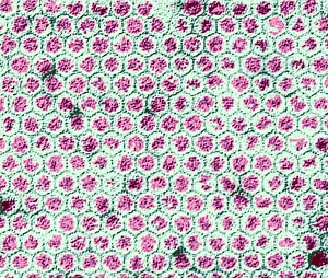
Polio virus. Coloured transmission electron micrograph (TEM) of a section through a para- crystalline array of polio virus particles

Background imageElectron Micrograph Collection: Herpes simplex virus infection, TEM

Herpes simplex virus infection, TEM
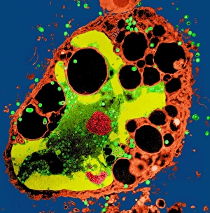
Herpes simplex virus infection. Coloured transmission electron micrograph (TEM) of a section through a cell being destroyed by herpes simplex virus infection

Background imageElectron Micrograph Collection: Adeno-associated virus, TEM

Adeno-associated virus, TEM
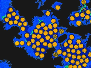
Adeno-associated virus. Coloured transmission electron micrograph (TEM) of particles of an unidentified adeno-associated virus (aV, green). A helper adenovirus (orange) is also seen

Background imageElectron Micrograph Collection: Yellow fever virus particles, TEM

Yellow fever virus particles, TEM
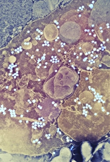
Yellow fever virus particles. Coloured transmission electron micrograph (TEM) of yellow fever virus particles (white) growing in culture


All Professionally Made to Order for Quick Shipping

"Unlocking the Hidden World: Exploring Electron Micrographs" Delve into the microscopic realm and witness the intricate beauty of life through electron micrographs. Discosphaera tubifera, coccolithophore: Marvel at the stunning calcium carbonate plates adorning this marine phytoplankton, resembling a delicate work of art. Crysotile asbestos: Peer into the dangerous fibers that make up this mineral, revealing its hazardous nature when inhaled. Liver: Journey inside this vital organ and observe its complex network of cells, unveiling its role in detoxification and metabolism. Cimex lectularius, bed bug: Get up close with these notorious pests as their exoskeletons reveal their resilience to survive even against our best efforts. Coloured TEM of Yersinia pestis bacteria: Witness the haunting beauty of these deadly bacteria responsible for causing plague outbreaks throughout history. Taraxacum officinale, dandelion (fruiting head): Explore the intricate structure of a dandelion's fruiting head under high magnification, showcasing nature's ingenious method for seed dispersal. Simulium damnosum, Simulian blackfly: Encounter these tiny insects known for transmitting river blindness as you uncover their detailed anatomy and feeding mechanisms. Norovirus particles, TEM: Enter the world of viruses as you observe norovirus particles - a common cause of gastrointestinal illness - providing insights into their structure and potential vulnerabilities for future treatments. 9 & 10 E. coli bacterium/bacteria : Dive deep into both individual E. coli cells or colonies to understand their role in digestion while also highlighting concerns surrounding foodborne illnesses caused by certain strains. Snail teeth : Discover how snails possess an unexpected weapon – razor-sharp teeth – enabling them to feed on tough plant material with ease; an evolutionary marvel. Chloroplast in cell of pea plant.