Malaria parasite, TEM

![]()
Wall Art and Photo Gifts from Science Photo Library
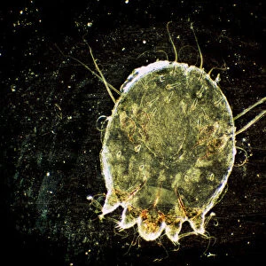
Malaria parasite, TEM
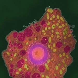
Malaria parasite. Image 4 of 10. Coloured transmission electron micrograph (TEM) of a sexual male malaria (Plasmodium sp.) microgametocyte in a mosquito (Anopheles sp.) gut. The microgametocyte releases male microgametes (purple, one at top right) after it is ingested by a mosquito feeding on an infected human. Each motile gamete possesses a flagellum built from fibrils (purple, paired) assembled near the gametocyte membrane. The male gametes fertilise the female macrogametocytes (not seen). Asexual reproduction then produces the stage that infects humans. Magnification: x5700 at 6x7cm size. For the malaria life cycle, see images M210/197-206
Science Photo Library features Science and Medical images including photos and illustrations
Media ID 6414796
© LSHTM/SCIENCE PHOTO LIBRARY
Assembling Assembly Electron Micrograph Fibril Fibrils Formation Forming Gamete Gametes Gametocyte Horizontal Infected Infection Infectious Life Cycle Malaria Malarial Micro Organism Microbe Microbiology Mosquito Parasite Parasites Parasitic Pathogenic Pathogens Production Protozoa Protozoan Release Reproducing Reproduction Sexual Stage Tissue Transmission Vector Borne Pathogen Section Sectioned
EDITORS COMMENTS
This print showcases a coloured transmission electron micrograph (TEM) of a male malaria parasite, specifically a microgametocyte, within the gut of a mosquito. The intricate details captured in this image provide valuable insights into the complex life cycle of the malaria parasite. The microgametocyte can be seen releasing male microgametes, represented by vibrant purple structures at the top right corner. These gametes possess flagella constructed from paired fibrils near the surface membrane of the gametocyte. It is through these motile gametes that fertilization occurs with female macrogametocytes, which unfortunately are not visible in this particular image. Upon ingestion by an Anopheles mosquito during its blood meal on an infected human, these male gametes play a crucial role in transmitting and perpetuating malaria infection. Once inside the mosquito's gut, they undergo further development and eventually give rise to stages capable of infecting humans again. With a magnification level of x5700 at 6x7cm size, this TEM image offers remarkable clarity and detail for studying various aspects of malarial parasites' biology and their interactions with their insect vectors. By understanding these processes at such microscopic levels, scientists gain deeper knowledge about pathogenic organisms like Plasmodium sp. , aiding efforts to develop effective strategies for combating this devastating disease.
MADE IN AUSTRALIA
Safe Shipping with 30 Day Money Back Guarantee
FREE PERSONALISATION*
We are proud to offer a range of customisation features including Personalised Captions, Color Filters and Picture Zoom Tools
SECURE PAYMENTS
We happily accept a wide range of payment options so you can pay for the things you need in the way that is most convenient for you
* Options may vary by product and licensing agreement. Zoomed Pictures can be adjusted in the Cart.