Fraud Blocker Skip to main content

Pathogen Collection

"Unseen Threats: Exploring the World of Pathogens" Budding yeast cell: A microscopic powerhouse, capable of causing infections in humans and animals alike

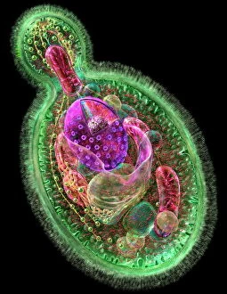
Background imagePathogen Collection: Budding yeast cell

Budding yeast cell. Computer artwork of asection through a yeast ( Candida albicans )cell that is reproducing asexually. A daughtercell (top left) is budding from the parent cell

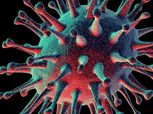
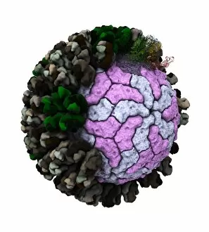
Background imagePathogen Collection: Avian flu virus

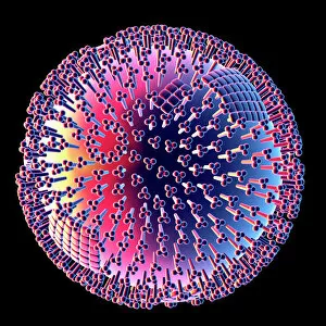
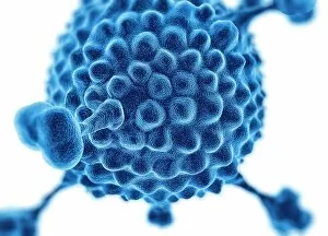
Avian flu virus, computer artwork. A virus is a tiny pathogenic particle comprising genetic material enclosed in a protein coat. The coat contains surface proteins (spikes)

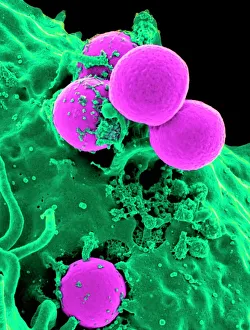
Background imagePathogen Collection: Neutrophil engulfing MRSA, SEM C018 / 8596

Neutrophil engulfing MRSA, SEM C018 / 8596
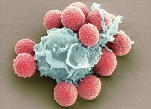
Neutrophil engulfing MRSA. Coloured scanning electron micrograph (SEM) of a neutrophil white blood cell (green) engulfing methicillin-resistant Staphylococcus aureus bacteria (MRSA, pink)

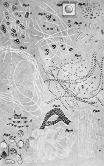
Background imagePathogen Collection: Anthrax cultures, historical diagram

Anthrax cultures, historical diagram
Anthrax cultures. 1876 diagram of Bacillus anthracis (Anthrax) bacteria cultured by Robert Koch. Along with Louis Pasteur, Koch is considered the founder of modern medical bacteriology

Background imagePathogen Collection: E. coli bacteria, SEM

E. coli bacteria, SEM
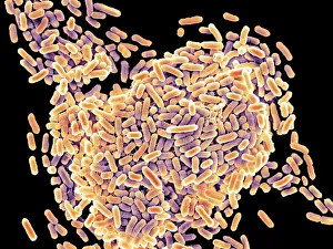
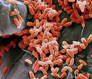
E. coli bacteria. Coloured scanning electron micrograph (SEM) of Escherichia coli bacteria. These bacteria are a normal part of the intestinal flora in humans and other animals

Background imagePathogen Collection: Herpes virus replicating

Herpes virus replicating, computer artwork.Viruses are only able to replicate in a host cell.The glycoprotein spikes in the virus envelope(green)

Background imagePathogen Collection: Salmonella bacteria, SEM

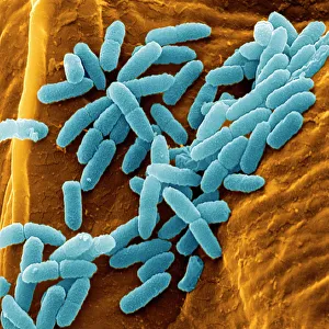
Salmonella bacteria, SEM
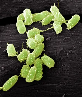
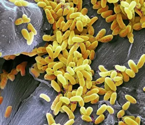
Salmonella bacteria, coloured scanning electron micrograph (SEM). These Gram-negative rod-shaped bacteria can cause food poisoning when eaten in contaminated food

Background imagePathogen Collection: Candida fungus, SEM

Candida fungus, SEM
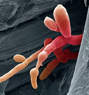
Candida fungus. Coloured scanning electron micrograph (SEM) of budding threads (hyphae) of a Candida fungus. The budding areas are where asexual reproduction is producing new fungus cells

Background imagePathogen Collection: HIV particle

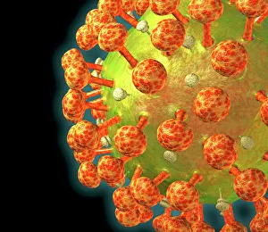
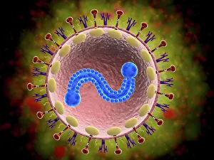
HIV particle, computer artwork. HIV (human immunodeficiency virus) causes AIDS (acquired immune deficiency syndrome). The virus consists of an RNA genome

Background imagePathogen Collection: Avian flu virus

Avian flu virus, computer artwork. A virus is a tiny pathogenic particle comprising genetic material enclosed in a protein coat. The coat contains surface proteins (spikes)

Background imagePathogen Collection: Norovirus particles, TEM

Norovirus particles, TEM
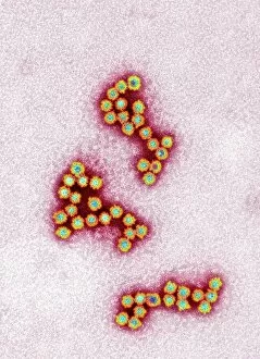
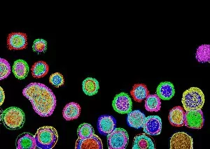
Norovirus particles. Coloured transmission electron micrograph (TEM) of norovirus particles. Norovirus is a genus of RNA (ribonucleic acid)

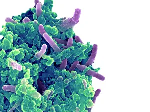
Background imagePathogen Collection: Tuberculosis bacteria

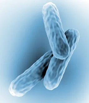
Tuberculosis bacteria. Computer artwork of Mycobacterium tuberculosis bacteria. These Gram- positive rod-shaped bacteria cause the disease tuberculosis

Background imagePathogen Collection: Pseudomonas aeruginosa bacteria, SEM

Pseudomonas aeruginosa bacteria, SEM
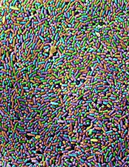
Pseudomonas aeruginosa bacteria, coloured scanning electron micrograph (SEM). These Gram-negative rod-shaped bacteria are found in soil and water, and as normal flora in the human intestine

Background imagePathogen Collection: Coloured TEM of a Salmonella bacterium

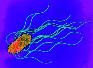
Coloured TEM of a Salmonella bacterium
Salmonella sp. Coloured transmission electron micrograph (TEM) of a Salmonella sp. bacterium. The cell is rod-shaped with long hair-like flagellae

Background imagePathogen Collection: Bacteria infecting a macrophage, SEM

Bacteria infecting a macrophage, SEM
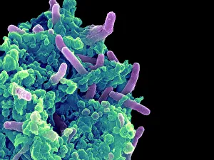
Bacteria infecting a macrophage. Coloured scanning electron micrograph (SEM) of Mycobacterium tuberculosis bacteria (purple) infecting a macrophage white blood cell

Background imagePathogen Collection: E. coli bacteria, SEM

E. coli bacteria, SEM
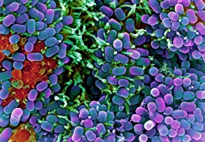
E. coli bacteria. Coloured scanning electron micrograph (SEM) of Escherichia coli bacteria (purple) taken from the small intestine of a child. E

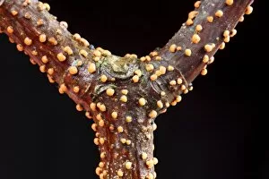
Background imagePathogen Collection: Coral Spot Fungus (Nectria cinnabarina) fruiting bodies, growing on Sycamore (Acer pseudoplatanus)

Coral Spot Fungus (Nectria cinnabarina) fruiting bodies, growing on Sycamore (Acer pseudoplatanus) twig, Powys, Wales, March

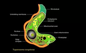
Background imagePathogen Collection: Trypanosome protozoan, artwork

Trypanosome protozoan, artwork
Trypanosome protozoan. Artwork of a section through a Trypanosoma congolense protozoan. This parasite is a cause of the disease nagana in cattle and other livestock in Africa

Background imagePathogen Collection: Salmonella bacteria, SEM

Salmonella bacteria, SEM
Salmonella bacteria, coloured scanning electron micrograph (SEM). Each rod-shaped structure is an individual bacterium. These bacteria occur mainly in human and animal intestines

Background imagePathogen Collection: Pseudomonas aeruginosa bacteria, SEM

Pseudomonas aeruginosa bacteria, SEM
Pseudomonas aeruginosa bacteria, coloured scanning electron micrograph (SEM). These Gram-negative rod-shaped bacteria are found in soil and water, and as normal flora in the human intestine

Background imagePathogen Collection: E. coli bacterium, TEM

E. coli bacterium, TEM
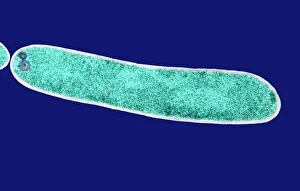
E. coli bacterium, coloured transmission electron micrograph (TEM). E. coli are Gram-negative rod-shaped bacteria that are part of the normal flora of the human gut

Background imagePathogen Collection: Macrophage engulfing TB bacteria, SEM

Macrophage engulfing TB bacteria, SEM
Macrophage engulfing TB bacteria. Coloured scanning electron micrograph (SEM) of a macrophage white blood cell (purple) engulfing a tuberculosis (Mycobacterium tuberculosis) bacterium (pink)

Background imagePathogen Collection: Microscopic view of human respiratory syncytial virus

Microscopic view of human respiratory syncytial virus (RSV). RSV causes respiratory tract infection of the lungs and breathing passages

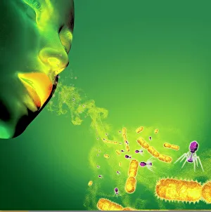
Background imagePathogen Collection: Infections spread by sneezing, artwork C013 / 5949

Infections spread by sneezing, artwork C013 / 5949
Infections spread by sneezing. Computer artwork showing virus particles (virions, blue) and bacteria (rod-shaped) in the spray produced by someone when they sneeze

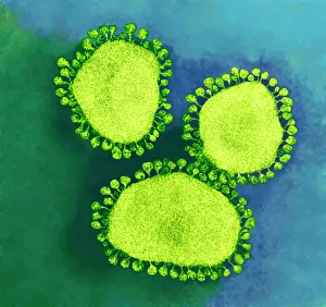
Background imagePathogen Collection: Coronavirus particles, TEM

Coronavirus particles, TEM
Coronavirus particles. Coloured transmission electron micrograph (TEM) of three coronavirus particles. Different strains of coronavirus are responsible for diseases such as the common cold

Background imagePathogen Collection: Illustration of response to infection after vaccination, involving microbe, antigens, antibody, plas

Illustration of response to infection after vaccination, involving microbe, antigens, antibody, plas

Background imagePathogen Collection: Microscopic view of chlamydia

Microscopic view of chlamydia. Chlamydia is a common sexually transmitted disease (STD) caused by a bacterium

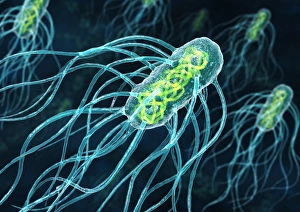
Background imagePathogen Collection: Salmonella bacteria, artwork C013 / 8818

Salmonella bacteria, artwork C013 / 8818
Salmonella bacteria, computer artwork. Salmonella sp. bacteria are gram-negative rod-shaped bacteria that have flagella (hair-like structures) that they use for locomotion

Background imagePathogen Collection: Phagocytosis of fungal spores, SEM

Phagocytosis of fungal spores, SEM
Phagocytosis of a fungus spore. Coloured scanning electron micrograph (SEM) of a neutrophil (blue) phagocytosing (engulfing and destroying) spores from the fungus Aspergillus fumigatus (red)

Background imagePathogen Collection: Influenza virus, computer artwork

Influenza virus, computer artwork
Influenza virus. Computer artwork of an influenza (flu) virus. The virus consists of a core of RNA (ribonucleic acid) genetic material surrounded by a protein coat

Background imagePathogen Collection: Influenza virus particles, TEM

Influenza virus particles, TEM
Influenza virus particles, coloured transmission electron micrograph (TEM). This virus is the cause of the respiratory disease influenza (flu) in humans and other animals

Background imagePathogen Collection: Hepatitis C viruses, TEM

Hepatitis C viruses, TEM
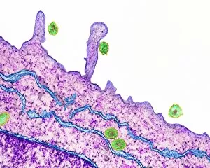
Hepatitis C viruses. Coloured transmission electron micrograph (TEM) of hepatitis C virus particles (green) infecting cultured liver cells (purple)

Background imagePathogen Collection: Pseudomonas aeruginosa bacteria, SEM

Pseudomonas aeruginosa bacteria, SEM
Pseudomonas aeruginosa bacteria, coloured scanning electron micrograph (SEM). These Gram-negative rod-shaped bacteria are found in soil and water, and as normal flora in the human intestine

Background imagePathogen Collection: Adenovirus, artwork

Adenovirus, artwork
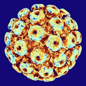
Adenovirus. Computer artwork of an adenovirus, showing the surface structure of the viruss outer protein coat (capsid). Adenoviruses are known to infect humans

Background imagePathogen Collection: Polyoma BK virus, artwork C013 / 7465

Polyoma BK virus, artwork C013 / 7465
Computer artwork of the capsid of a polyoma BK virus. This polyomavirus is common in the urinary tract of adults, where it lives without harming its host

Background imagePathogen Collection: Salmonella culture

Salmonella culture
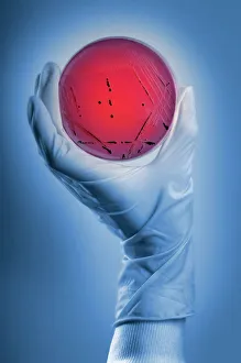
MODEL RELEASED. Salmonella culture. Microbiologist holding a petri dish containing a culture of Salmonella sp. bacteria. These bacteria are the cause of salmonellosis (food poisoning) in humans

Background imagePathogen Collection: Mouse malaria parasite, SEM

Mouse malaria parasite, SEM
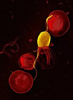
Mouse malaria parasite. Coloured scanning electron micrograph (SEM) of a Plasmodium berghei protozoan (yellow) and red blood cells. P. berghei is the parasite that causes malaria in mice

Background imagePathogen Collection: Rift Valley fever virus, TEM

Rift Valley fever virus, TEM

Background imagePathogen Collection: Vesicular stomatitis virus, TEM

Vesicular stomatitis virus, TEM
Vesicular stomatitis virus. Coloured transmission electron micrograph (TEM) of vesicular stomatitis virus (VSV) particles (blue) budding from a host cell (red)

Background imagePathogen Collection: Paramyxovirus particles, TEM

Paramyxovirus particles, TEM
Sendai virus. Coloured transmission electron micrograph (TEM) of Sendai virus particles (virions, orange). The protein coat (capsid) of one of the particles has split

Background imagePathogen Collection: Mycobacterium chelonae bacteria, SEM

Mycobacterium chelonae bacteria, SEM
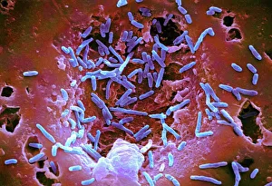
Mycobacterium chelonae bacteria, coloured scanning electron micrograph (SEM). These Gram-positive rod-shaped bacteria are found in soil and water

Background imagePathogen Collection: E. coli bacterium, TEM

E. coli bacterium, TEM
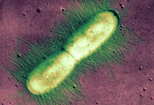
E. coli bacterium. Coloured transmission electron micrograph (TEM) of an Escherichia coli bacterium in the early stages of binary fission, the process by which the bacterium divides

Background imagePathogen Collection: Cholera bacteria, artwork

Cholera bacteria, artwork
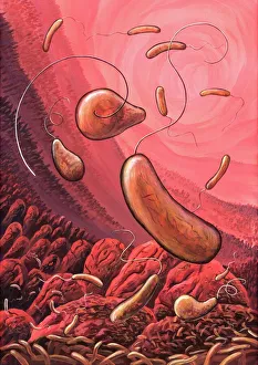
Cholera bacteria. Artwork of Vibrio cholerae bacteria in the small intestine. These Gram-negative rod-shaped bacteria have a single polar flagellum (tail-like structure)

Background imagePathogen Collection: Rotavirus particle, artwork

Rotavirus particle, artwork
Rotavirus particle. Cut-away artwork showing the structure of the rotavirus icosahedral capsid (protein coat). The capsid, which encloses the RNA (ribonucleic acid) genome

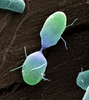
Background imagePathogen Collection: Salmonella bacterium dividing, SEM

Salmonella bacterium dividing, SEM
Salmonella bacterium dividing. Coloured scanning electron micrograph (SEM) of of a Salmonella bacterium dividing. The two new daughter cells are seen at upper right and lower left

Background imagePathogen Collection: Bacteria infecting a macrophage, SEM

Bacteria infecting a macrophage, SEM
Bacteria infecting a macrophage. Coloured scanning electron micrograph (SEM) of Mycobacterium tuberculosis bacteria (purple) infecting a macrophage white blood cell

Background imagePathogen Collection: An engraving depicting Hoof fungus (Fomes fomentarius), the source of German tinder or touchwood

An engraving depicting Hoof fungus (Fomes fomentarius), the source of German tinder or touchwood
5311405 An engraving depicting Hoof fungus (Fomes fomentarius), the source of German tinder or touchwood. Specimens were left to grow to a large size so that the tinder was harvested

Background imagePathogen Collection: The Microbe (colour litho)

The Microbe (colour litho)
8633854 The Microbe (colour litho) by Thompson, George Henry (1853-1933); Private Collection; (add.info.: The Microbe. Illustration for The Poet and Other Animals by Harold Richardon (Ernest Nister)


All Professionally Made to Order for Quick Shipping

"Unseen Threats: Exploring the World of Pathogens" Budding yeast cell: A microscopic powerhouse, capable of causing infections in humans and animals alike. Avian flu virus: A notorious pathogen that poses a significant threat to both birds and humans, with potential for devastating global outbreaks. Neutrophil engulfing MRSA, SEM C018 / 8596: Witness the incredible defense mechanism of our immune system as it battles against antibiotic-resistant bacteria. E. Coli bacteria, SEM: Unveiling the minuscule culprits behind foodborne illnesses and urinary tract infections. Salmonella bacteria, SEM: Discover the intricate structure of this common bacterial pathogen responsible for causing severe gastrointestinal distress. Candida fungus, SEM: Peering into the world of fungal infections as Candida takes center stage in various ailments from thrush to systemic candidiasis. Coral Spot Fungus (Nectria cinnabarina) fruiting bodies on Sycamore twig, Powys, Wales: Marvel at nature's ability to harbor pathogens even within picturesque settings like these vibrant red fruiting bodies growing on trees. HIV particle: Delve into the complex realm of retroviruses with a focus on HIV/AIDS – an ongoing global health crisis demanding attention and research breakthroughs. Anthrax cultures, historical diagram: Tracing back through history to understand one of humanity's oldest known pathogens – anthrax – its deadly impact and potential bioweapon applications throughout time. Norovirus particles, TEM: Get up close with these tiny viral particles responsible for stomach flu outbreaks worldwide; their resilience challenges public health measures constantly. Tuberculosis bacteria - Explore the persistent Mycobacterium tuberculosis that causes tuberculosis disease affecting millions globally despite medical advancements Avian flu virus - Investigate avian influenza viruses' ability to jump species barriers, posing a constant threat to both birds and humans.