
Freeze Fractured Collection
"Exploring the Intricate World of Freeze-Fractured Baby Cream and Skin Structures" In the realm of scientific imaging


All Professionally Made to Order for Quick Shipping
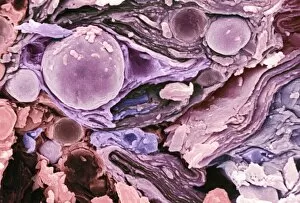
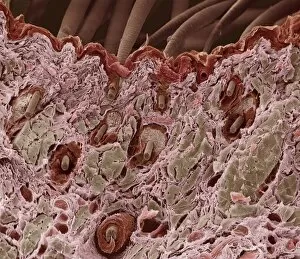
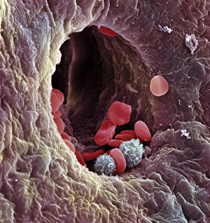
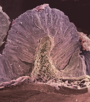
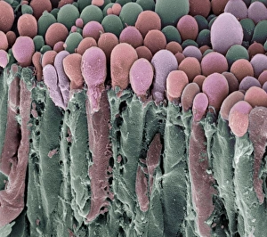
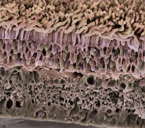
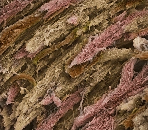
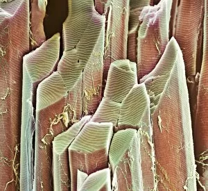
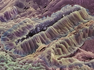
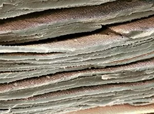
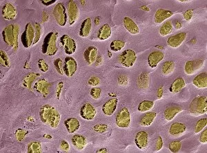
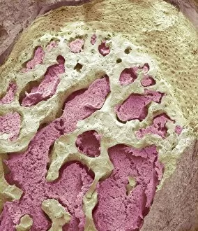
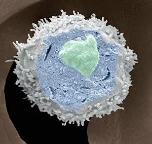
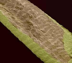
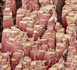
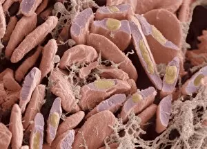
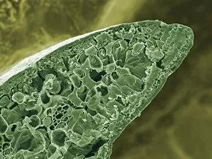
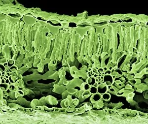
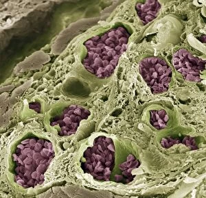
"Exploring the Intricate World of Freeze-Fractured Baby Cream and Skin Structures" In the realm of scientific imaging, freeze-fracturing offers a fascinating glimpse into the hidden intricacies of various substances and biological structures. Captured under scanning electron microscopy (SEM), these captivating images reveal remarkable details that are otherwise invisible to the naked eye. The first set of images, labeled as "Freeze-fractured baby cream, SEM C017 / 7133, " "Freeze-fractured baby cream, SEM C017 / 7134, " and "Freeze-fractured baby cream, SEM C017 / 7135, " take us on an enchanting journey through this unique product's composition. The frozen fractures expose its internal structure with mesmerizing patterns resembling delicate ice crystals or intricate lacework. These close-ups offer a rare opportunity to appreciate the complexity behind everyday items we often take for granted. Moving beyond skincare products, our exploration continues with stunning visuals captured under SEM. The image titled "Hair follicle, SEM" invites us into the microscopic world beneath our scalps. It unveils hair follicles in astonishing detail – their elongated shapes and distinct layers forming a tapestry that contributes to our individuality. Similarly, we encounter another series labeled as "Skin layers, SEM. " These captivating snapshots showcase different depths within our skin – from epidermis to dermis – each layer playing a crucial role in protecting and nurturing our bodies. Delicate ridges intertwine like miniature mountain ranges while cells appear like tiny building blocks constructing this incredible fortress called skin. Returning once again to hair-related wonders, we delve deeper into hair follicles through multiple images titled "Hair follicle, SEM. " Each photograph unravels new mysteries surrounding these vital structures responsible for hair growth. Their intricate arrangements resemble architectural marvels designed by nature itself.