
Gaseous Exchange Collection
"Gaseous Exchange: The Intricate Dance of Life" In this captivating engraving, we delve into the fascinating world within living organisms


All Professionally Made to Order for Quick Shipping
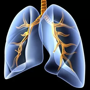
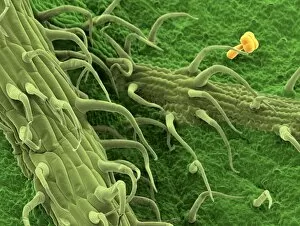
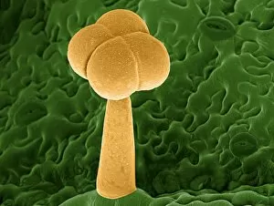
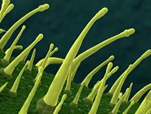
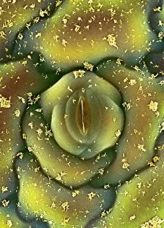
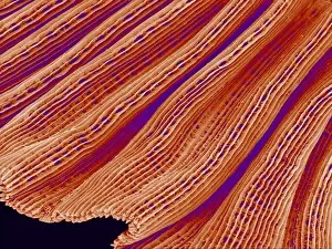
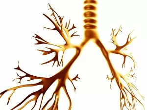
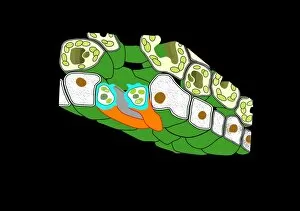
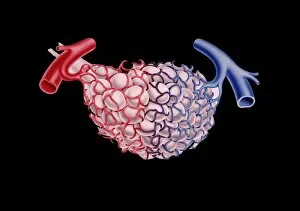
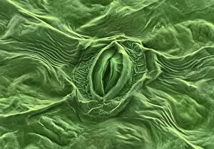
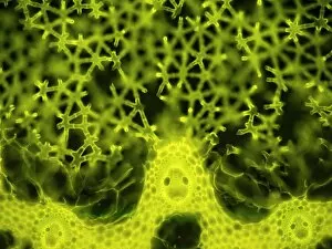
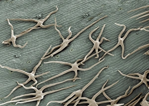
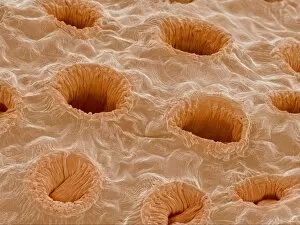
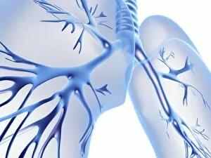
"Gaseous Exchange: The Intricate Dance of Life" In this captivating engraving, we delve into the fascinating world within living organisms. The intricate anatomy of organs comes to life, revealing the remarkable mechanisms that enable survival. Firstly, we witness a mesmerizing transformation as yeast and sugar solution ferment, producing a froth brimming with carbon dioxide through anaerobic respiration. This process highlights how even in the absence of oxygen, life finds ingenious ways to sustain itself. Moving on to the bronchial tree artwork series, we are transported into the realm of mammals' respiratory system. These branching passageways intricately connect our lungs to every corner of our bodies, ensuring efficient gas exchange. With each breath we take, oxygen fills our lungs while carbon dioxide is expelled – an essential dance that keeps us alive. But it's not just mammals who possess such incredible adaptations; fish too have their own unique method for gaseous exchange. Through scanning electron microscopy (SEM), we explore fish gills in astonishing detail. These delicate structures allow water rich in oxygen to flow over them while extracting precious O2 molecules and releasing CO2 back into their watery environment. The enchanting images captured by SEM C015/8613/8663/8661/8660 showcase nature's ingenuity at its finest – a testament to evolution's ceaseless quest for survival strategies. Gaseous exchange serves as a reminder that life is interconnected and constantly adapting to its surroundings. Whether it be through intricate anatomical engravings or microscopic wonders revealed under SEMs lens - these snapshots offer glimpses into the awe-inspiring beauty hidden within every breath taken by living organisms around us.