
Gastric Collection
"Gastric: Exploring the Intricate World of Digestion and Health" Step into the fascinating realm of the human digestive system, where art meets science


All Professionally Made to Order for Quick Shipping
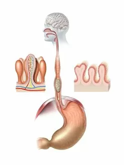
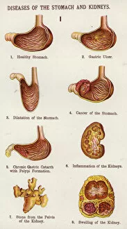
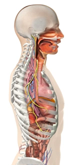
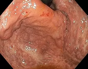
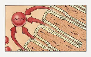
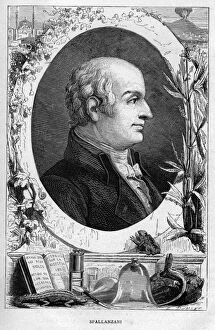
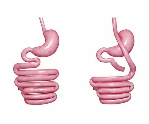
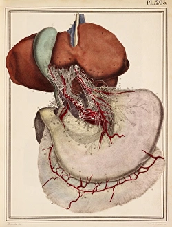
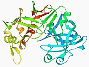
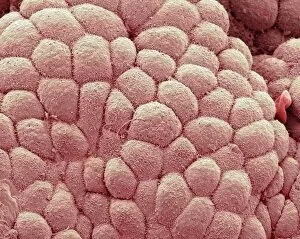
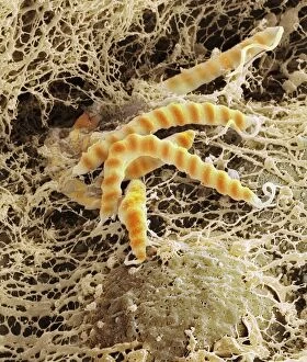
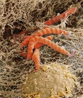
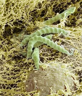
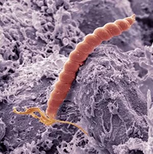
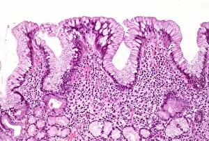
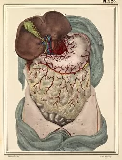
"Gastric: Exploring the Intricate World of Digestion and Health" Step into the fascinating realm of the human digestive system, where art meets science. From intricate artwork depicting diseases of the stomach and kidneys to historical illustrations showcasing medical breakthroughs, this captivating journey will leave you in awe. Delve into a world plagued by typhus as you witness haunting images of sufferers in Kniagin, Russia, being transported on trains. Discover the connection between gastric health and our nervous system through stunning artwork highlighting the anatomy of the vagus nerve. Uncover rare conditions like gastric antral vascular ectasia with vivid visuals that shed light on its impact. Marvel at Rudolph Valentino's timeless charm as he poses alongside his beloved dogs in 1926. Witness how nature's wonders work within us through detailed illustrations showcasing gastric glands secreting pepsin to break down proteins into digestible peptides. Transport yourself back to 19th-century France with a line engraving capturing human stomach glands meticulously producing essential gastric juices. Learn about Lazzaro Spallanzani, an Italian biologist whose groundbreaking research challenged prevailing beliefs about bacteria. Explore innovative solutions like gastric bypass depicted through thought-provoking artworks that revolutionize weight loss treatments. Join us on this enlightening journey as we unravel mysteries surrounding our most vital organ - the stomach. Gain a deeper understanding of digestion, health, and scientific advancements that continue to shape our understanding of gastronomy today.