
Gastroenterological Collection
"Gastroenterological Wonders: Exploring the Intricacies of the Digestive System" Delicate and captivating


All Professionally Made to Order for Quick Shipping
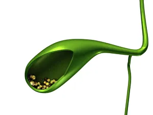
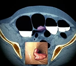
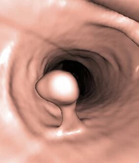
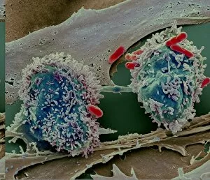
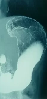
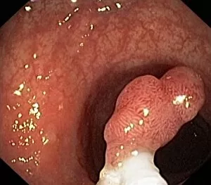
"Gastroenterological Wonders: Exploring the Intricacies of the Digestive System" Delicate and captivating, this artwork beautifully depicts the formation of gallstones within the gallbladder. Dive into a microscopic world with this stunning SEM image showcasing colorectal cancer cells, reminding us of the importance of early detection and prevention. A mesmerizing light micrograph reveals the intricate details of gastritis, highlighting its impact on our digestive system's delicate lining. Witnessing threadworms in their natural habitat through an SEM image provides valuable insights into these parasitic creatures' presence within our gut. Another glimpse into gastritis through a captivating light micrograph emphasizes its diverse manifestations and effects on our gastrointestinal health. Colitis comes to life under a microscope as this vibrant light micrograph showcases inflamed tissue, urging us to prioritize proper management and care for this condition. Ischaemic bowel takes center stage in this enlightening light micrograph, shedding light on a potentially serious condition that demands immediate medical attention. Threadworms make another appearance in yet another striking SEM image, serving as a reminder to stay vigilant against these common intestinal parasites. Intriguingly complex and visually compelling, gastroenterology unravels mysteries hidden deep within our bodies—reminding us to nurture our digestive health for overall well-being.