
Goblet Cell Collection
"Goblet cells: Unveiling the Secretive Guardians of Our Respiratory and Digestive Systems" In the intricate world of our respiratory and digestive systems


All Professionally Made to Order for Quick Shipping
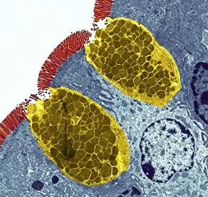
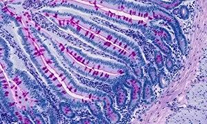
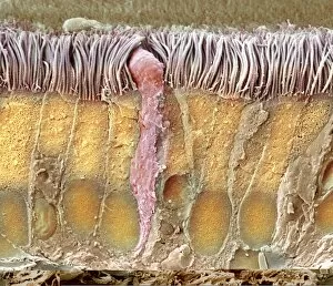
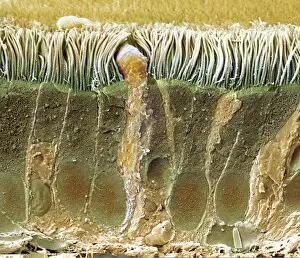
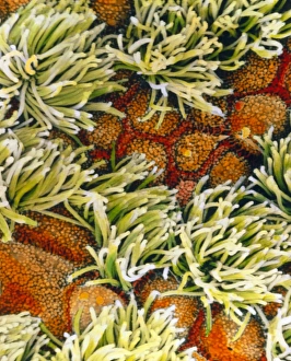
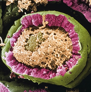
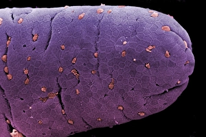
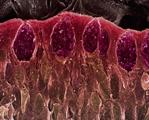
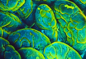
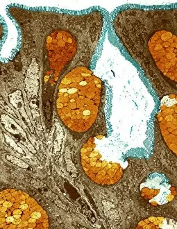
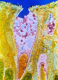
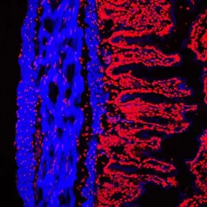
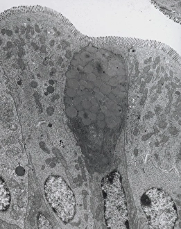
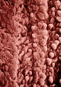
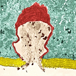
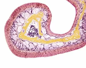
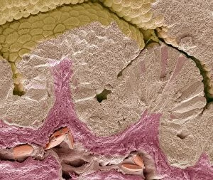
"Goblet cells: Unveiling the Secretive Guardians of Our Respiratory and Digestive Systems" In the intricate world of our respiratory and digestive systems, there exists a remarkable cell type known as goblet cells. These unique cells play a crucial role in maintaining the health and functionality of our trachea, small intestine, and colon. Underneath the lens of scanning electron microscopy (SEM), we witness the mesmerizing beauty of these goblet cells lining the trachea. SEM images C013/7126, C013/7122, and C013/7127 reveal their distinctive shape - resembling an elegant goblet or wine glass. Their elongated bodies are adorned with tiny hair-like projections called cilia that aid in sweeping away harmful particles from our airways. Moving deeper into this microscopic journey, we encounter another stunning image captured by SEM showcasing the trachea's overall structure. This captivating view highlights how goblet cells work harmoniously alongside other specialized cells to create a protective barrier against foreign invaders. Shifting gears to light micrographs, we delve into different sections of our digestive system where goblet cells also hold significant importance. In one such image taken from the small intestine, these incredible guardians can be seen scattered throughout its lining (light micrograph). They stand tall amidst absorptive epithelial cells like sentinels guarding against pathogens while secreting mucus to lubricate food passage. Venturing further down into our colon through yet another light micrograph F006/9806 reveals an abundance of goblet cells at work. Here they form clusters within crypts on top of columnar epithelial tissue - their presence essential for maintaining proper gut function by facilitating smooth waste elimination. To truly appreciate their complexity up close, transmission electron microscopy (TEM) provides us with detailed insights into individual goblet cell structures. TEM imagery showcases their secretory granules filled with mucins – proteins responsible for producing the protective mucus layer that shields our delicate tissues from harm.