
Gram Positive Bacteria Collection
Gram positive bacteria are a diverse group of microorganisms that play a significant role in both human health and disease


All Professionally Made to Order for Quick Shipping
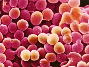
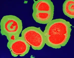
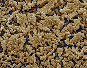
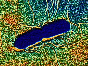
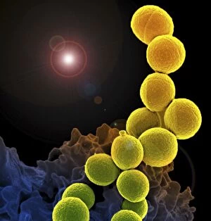
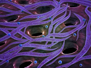
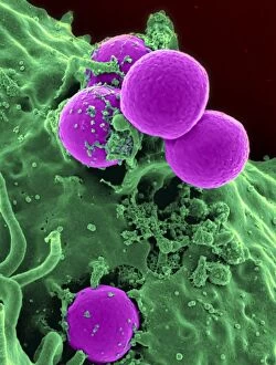
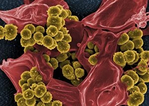
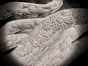
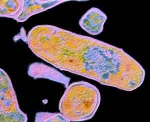
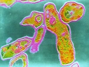
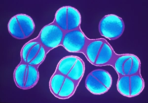
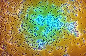
Gram positive bacteria are a diverse group of microorganisms that play a significant role in both human health and disease. One well-known example is Staphylococcus aureus, which can cause various infections ranging from skin infections to life-threatening conditions. Another notable member is MRSA resistant Staphylococcus bacteria, which poses a challenge due to its resistance to common antibiotics. Illustrations of Streptococcus bacteria showcase their characteristic chain-like arrangement, responsible for causing strep throat and other respiratory infections. Bacillus subtilis bacteria, as seen through scanning electron microscopy (SEM), display their rod-shaped structure and are commonly found in soil. Microscopic views of staphylococcus reveal their spherical shape arranged in clusters or pairs. These gram-positive cocci can cause numerous diseases such as skin abscesses and bloodstream infections. Similarly, diplococcus bacterium appears under the microscope as two round cells joined together, often associated with certain types of pneumonia. Listeria monocytogenes, another gram-positive bacterium depicted at the microscopic level, is known for causing foodborne illnesses that can be particularly dangerous for pregnant women and individuals with weakened immune systems. In diagnostic settings, red blood cells on an agar plate serve as indicators for infection caused by gram-positive bacteria. By observing bacterial growth patterns around these cells, healthcare professionals can identify specific pathogens present in patient samples. Lastly but not least important is Methicillin-resistant Staphylococcus aureus (MRSA), shown under the microscope with its distinctive appearance. This antibiotic-resistant strain has become a major concern worldwide due to its ability to cause severe hospital-acquired infections.