Fraud Blocker Skip to main content

Hair Shaft Collection

The hair shaft, an intricate part of our body, is a fascinating subject to explore

Background imageHair Shaft Collection: Coloured SEM of a human hair on the skin

Coloured SEM of a human hair on the skin
Hair on skin. Coloured Scanning Electron Micro- graph (SEM) of a hair on the surface of human skin. At centre is a shaft of hair (dark grey) which has emerged from a follicle in the skin

Background imageHair Shaft Collection: Coloured SEM of a section through a hair follicle

Coloured SEM of a section through a hair follicle
Hair follicle. Coloured scanning electron micrograph (SEM) of a cross-section through a hair follicle below the skin surface

Background imageHair Shaft Collection: Lymphocytes in hair follicle, SEM

Lymphocytes in hair follicle, SEM
Lymphocytes in hair follicle, coloured scanning electron micrograph (SEM). Large numbers of lymphocytes (yellow) are seen in inflamed and necrotic tissue at the base of hair shafts (red)

Background imageHair Shaft Collection: Daubentons bat hairs, SEM

Daubentons bat hairs, SEM
Bat hairs. Coloured scanning electron micrograph (SEM) of hairs from Daubentons bat (Myotis daubentonii). Each hair shaft has an outer cuticle made up of overlapping scales, or plates

Background imageHair Shaft Collection: SEM of bat hair

SEM of bat hair
Bat hairs. Coloured scanning electron micrograph (SEM) of shafts of hair from Daubentons bat, Myotis daubentoni. Each hair shaft has an outer cuticle made up of overlapping scales or plates

Background imageHair Shaft Collection: Damaged human hair shaft SEM

Damaged human hair shaft SEM
Damaged human hair shaft, coloured scanning electron micrograph (SEM). This can be caused by overheating the hair using curling tongs or hair straigheners, as well as chemical dyes

Background imageHair Shaft Collection: Human hair shaft with split ends SEM

Human hair shaft with split ends SEM
Human hair shaft with split ends, coloured scanning electron micrograph (SEM). This can be caused by overheating the hair using curling tongs or hair straigheners, as well as chemical dyes

Background imageHair Shaft Collection: SEM of hair on scalp

SEM of hair on scalp
Hair. Coloured scanning electron micrograph of hairs protruding from the surface of the scalp. Numerous desquamating cells (pink) are concentrically arranged around the base of the hair shaft

Background imageHair Shaft Collection: Human hair

Human hair
False-colour scanning electron micrograph (SEM) of a clean human hair. Hair is a fibrous protein (keratin) which develops out of a tubular follicle located in the epidermis of the skin

Background imageHair Shaft Collection: Pyoderma skin disease, SEM

Pyoderma skin disease, SEM
Pyoderma skin disease, coloured scanning electron micrograph (SEM). Large numbers of lymphocytes (white) and red blood cells are seen in necrotic tissue surrounding a hair shaft

Background imageHair Shaft Collection: Hair shaft anatomy, artwork

Hair shaft anatomy, artwork
Hair shaft anatomy. Cutaway artwork showing the internal structure and anatomy of a human hair. Hair is made of a fibrous protein called keratin

Background imageHair Shaft Collection: Bacterial skin infections, artwork

Bacterial skin infections, artwork
Bacterial skin infections, computer artwork. Round (green, cocci) and cylindrical (blue, bacilli) bacteria on skin, with hair shafts also seen

Background imageHair Shaft Collection: Lymphocytes in skin disorder, SEM

Lymphocytes in skin disorder, SEM
Lymphocytes in skin disorder, coloured scanning electron micrograph (SEM). Lymphocytes (light green) are seen in inflamed and necrotic tissue


All Professionally Made to Order for Quick Shipping

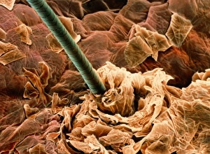
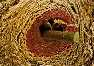
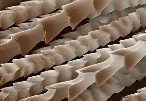
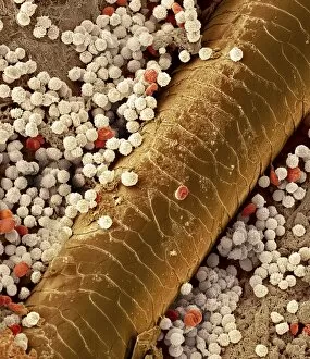
The hair shaft, an intricate part of our body, is a fascinating subject to explore. From the coloured scanning electron microscope (SEM) images capturing the beauty of human hair on the skin and sections through hair follicles, to the surprising presence of lymphocytes within these follicles, there is much to discover. Biomedical illustrations provide us with a cross-sectional view of the structure of our skin where these hair shafts reside. But it's not just humans that possess intriguing hairs – Daubenton's bat hairs also captivate us under SEM. Examining damaged human hair shafts reveals their vulnerability and split ends, reminding us to care for them properly. The diversity in human hairs showcased by SEM highlights their unique characteristics and textures. Zooming in further, we observe scalp hairs up close – each strand telling its own story. Finally, exploring pyoderma skin disease through SEM allows us to understand how this condition affects both our skin and its associated hair follicles.