
Histopathological Collection
Histopathological investigations offer valuable insights into various diseases and conditions affecting the human body


All Professionally Made to Order for Quick Shipping
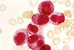
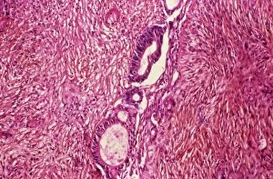
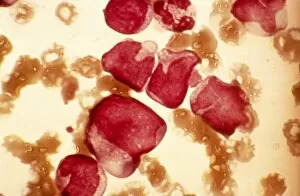
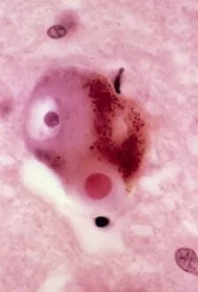
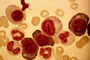
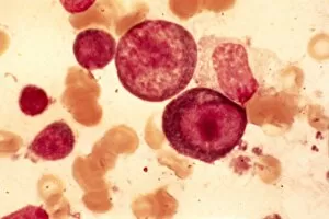
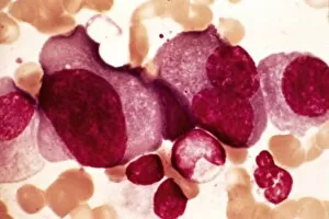
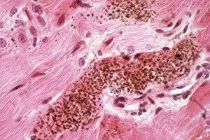
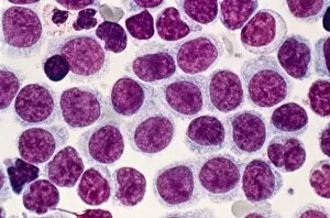
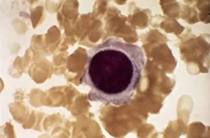
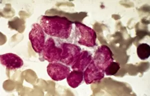
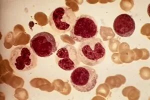
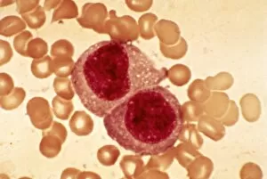
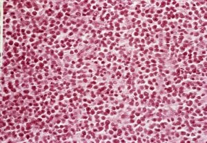
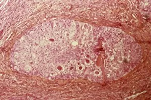
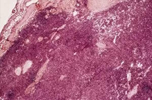
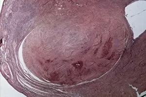
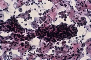
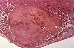
Histopathological investigations offer valuable insights into various diseases and conditions affecting the human body. From identifying specific cellular abnormalities to understanding the progression of life-threatening illnesses, histopathology plays a crucial role in diagnosis and treatment. Dohle bodies in blood cells, as seen through a micrograph, provide essential clues for medical professionals studying infections or inflammatory disorders. These distinctive blue-grey structures within neutrophils can indicate underlying health issues. In another micrograph, acute promyelocytic leukemia is evident with its characteristic abnormal promyelocytes. This aggressive form of leukemia requires prompt identification for effective management. A light micrograph showcasing ovarian cancer (C015 / 7103) highlights the importance of early detection in combating this deadly disease that affects women worldwide. Cystic fibrosis, a genetic disorder primarily affecting the respiratory system, can be identified through histopathological examinations revealing thick mucus accumulation and damaged lung tissue. Gout crystals are easily recognizable under microscopic examination due to their needle-like appearance. The presence of these crystals helps confirm gout as the cause behind painful joint inflammation. Examining lymphocytes within hair follicles using scanning electron microscopy provides researchers with valuable data about immune responses occurring at localized sites on our skin. Biopsy slides (F007 / 0316) play an integral role in diagnosing various conditions by allowing pathologists to analyze tissue samples under high magnification. These slides aid in determining appropriate treatment plans tailored to each patient's needs. Light micrographs depicting testicular cancer highlight distinct cellular changes that help differentiate it from other forms of malignancies originating from testicular tissues. Dementia, characterized by cognitive decline and memory loss, leaves its mark on brain tissue visible through light microscopy. Identifying key pathological features aids in understanding this complex neurodegenerative condition better. Multiple myeloma manifests itself within bone marrow plasma cells and can be observed via light microscopy techniques. Accurate diagnosis enables healthcare professionals to devise targeted treatment strategies.