Fraud Blocker Skip to main content

Histopathological Collection (page 2)

Histopathological investigations offer valuable insights into various diseases and conditions affecting the human body

Background imageHistopathological Collection: Acute leukaemia, light micrograph

Acute leukaemia, light micrograph
Acute leukaemia. Light micrograph of blood cells from bone marrow in a case of acute leukaemia. These cells include a polynuclear degranulated basophil and myeloblasts

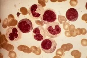
Background imageHistopathological Collection: Chronic myeloid leukaemia, micrograph

Chronic myeloid leukaemia, micrograph
Chronic myeloid leukaemia. Light micrograph of blood cells from bone marrow in a case of chronic myeloid leukaemia. Leukaemia is a cancer where certain blood cells form in excess

Background imageHistopathological Collection: Reticulosarcoma, light micrograph

Reticulosarcoma, light micrograph
Reticulosarcoma. Light micrograph of reticulocyte blood cells (red, one at upper left) from a lymphatic ganglion in a case of reticulosarcoma

Background imageHistopathological Collection: Langerhans cell histiocytosis, micrograph

Langerhans cell histiocytosis, micrograph
Langerhans cell histiocytosis. Light micrograph of blood cells in a case of Langerhans cell histiocytosis (LCH), also called X-type histiocytosis

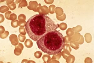
Background imageHistopathological Collection: Hodgkins disease, light micrograph

Hodgkins disease, light micrograph
Hodgkins disease. Light micrograph of two non-dystrophic histiocyte blood cells (centre) in a case of Hodgkins disease, also called Hodgkins lymphoma

Background imageHistopathological Collection: Reticulosarcoma, light micrograph C015 / 7133

Reticulosarcoma, light micrograph C015 / 7133
Reticulosarcoma. Light micrograph of reticulocyte blood cells (red, one at upper left) from a lymphatic ganglion in a case of reticulosarcoma

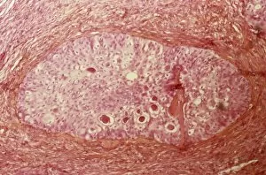
Background imageHistopathological Collection: Brenner tumour of the ovary C015 / 7137

Brenner tumour of the ovary C015 / 7137
Brenner tumour of the ovary. Light micrograph of a section through an ovary showing a Brenner tumour (centre). This rare benign (non-cancerous)

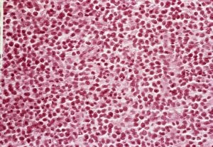
Background imageHistopathological Collection: Langerhans cell histiocytosis C015 / 7135

Langerhans cell histiocytosis C015 / 7135
Langerhans cell histiocytosis. Light micrograph of a section through a lymph node showing Langerhans cell histiocytosis (LCH), also known as granulomatosis

Background imageHistopathological Collection: Lung silicosis, light micrograph C015 / 7129

Lung silicosis, light micrograph C015 / 7129
Lung silicosis. Light micrograph of a section through a lung with silicosis, a type of pneumoconiosis caused by exposure to silica dusts. The lung tissue has become fibrous (centre)

Background imageHistopathological Collection: Thymus gland cancer, light micrograph C015 / 7128

Thymus gland cancer, light micrograph C015 / 7128
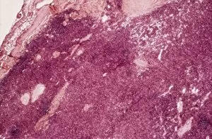
Thymus cancer. Light micrograph of a section through a thymus gland with thymoma cancer. The thymus is a gland in the upper chest that plays an important role in the immune system

Background imageHistopathological Collection: Endometrial hyperplasia, light micrograph C015 / 7104

Endometrial hyperplasia, light micrograph C015 / 7104
Endometrial hyperplasia. Light micrograph of a section through tissue from the endometrium, in a case of endometrial hyperplasia. The endometrium is the uterus lining

Background imageHistopathological Collection: Uterine fibroid, light micrograph C015 / 7101

Uterine fibroid, light micrograph C015 / 7101
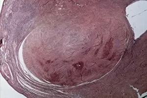
Uterine fibroid. Light micrograph of a section through a uterine fibroid (centre). A fibroid is a fibrous benign tumour originating from muscular tissue

Background imageHistopathological Collection: Cervical cancer, light micrograph C015 / 7000

Cervical cancer, light micrograph C015 / 7000
Cervical cancer. Light micrograph of a section through tissue from the cervix (neck of the womb) showing intraepithelial carcinoma (centre), a form of cancer

Background imageHistopathological Collection: Cervical cancer, light micrograph C015 / 6999

Cervical cancer, light micrograph C015 / 6999
Cervical cancer. Light micrograph of a section through tissue from the cervix (neck of the womb) showing intraepithelial carcinoma (across centre), a form of cancer

Background imageHistopathological Collection: Cervical cancer, light micrograph C015 / 6998

Cervical cancer, light micrograph C015 / 6998
Cervical cancer. Light micrograph of a section through tissue from the cervix (neck of the womb) showing intraepithelial carcinoma (dark areas), a form of cancer

Background imageHistopathological Collection: Cervical cancer, light micrograph C015 / 6997

Cervical cancer, light micrograph C015 / 6997
Cervical cancer. Light micrograph of a section through tissue from the cervix (neck of the womb) showing intraepithelial carcinoma (across centre), a form of cancer

Background imageHistopathological Collection: Cervical smear showing endometrial cancer C015 / 6750

Cervical smear showing endometrial cancer C015 / 6750
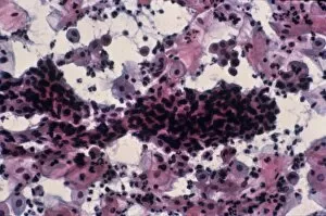
Cervical smear showing endometrial cancer. Light micrograph of cells from a cervical smear test showing endometrial sarcoma. This is a cancer of the lining of the womb, the endometrium

Background imageHistopathological Collection: Cervical wart, light micrograph C015 / 6747

Cervical wart, light micrograph C015 / 6747
Cervical wart. Light micrograph of a section through a wart (upper frame) on the cervix (neck of the uterus). This wart is caused by the human papilloma virus (HPV)

Background imageHistopathological Collection: Uterine fibroid, light micrograph C015 / 6737

Uterine fibroid, light micrograph C015 / 6737
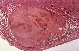
Uterine fibroid. Light micrograph of a section through a uterine fibroid (oval). A fibroid is a fibrous benign tumour originating from muscular tissue

Background imageHistopathological Collection: Cervical wart, light micrograph C015 / 6055

Cervical wart, light micrograph C015 / 6055
Cervical wart. Light micrograph of a section through a condyloma, an abnormal wart-like growth, on the cervix (neck of the uterus)

Background imageHistopathological Collection: Chronic myeloid leukaemia, micrograph C015 / 6227

Chronic myeloid leukaemia, micrograph C015 / 6227
Chronic myeloid leukaemia. Light micrograph of bone marrow from a patient with chronic myeloid leukaemia (CML). This cancer affects the myeloid tissue (bone marrow)

Background imageHistopathological Collection: Acute myeloid leukaemia C015 / 6225

Acute myeloid leukaemia C015 / 6225
Acute myeloid leukaemia. Light micrograph of bone marrow from a patient with acute myeloid leukaemia (AML). This cancer affects the myeloid tissue (bone marrow)

Background imageHistopathological Collection: Uterine fibroid, light micrograph C015 / 6413

Uterine fibroid, light micrograph C015 / 6413
Uterine fibroid. Light micrograph of a section through a uterine fibroid. A fibroid is a fibrous benign tumour originating from muscular tissue

Background imageHistopathological Collection: Uterine fibroid, light micrograph C015 / 6411

Uterine fibroid, light micrograph C015 / 6411
Uterine fibroid. Light micrograph of a section through a uterine fibroid (centre). A fibroid is a fibrous benign tumour originating from muscular tissue

Background imageHistopathological Collection: Uterine fibroid, light micrograph C015 / 6412

Uterine fibroid, light micrograph C015 / 6412
Uterine fibroid. Light micrograph of a section through a uterine fibroid (oval). A fibroid is a fibrous benign tumour originating from muscular tissue

Background imageHistopathological Collection: Paratubal cysts, light micrograph C015 / 6410

Paratubal cysts, light micrograph C015 / 6410
Paratubal cysts. Light micrograph of a section through a fallopian tube (oviduct) with three paratubal cysts (top). Cysts are fluid-filled sacs

Background imageHistopathological Collection: Cervical cyst, light micrograph C015 / 6408

Cervical cyst, light micrograph C015 / 6408
Cervical cyst. Light micrograph of a section through tissue from the cervix (neck of the womb) showing a mucous cyst (pale, centre)

Background imageHistopathological Collection: Listeriosis, light micrograph C015 / 6405

Listeriosis, light micrograph C015 / 6405
Listeriosis. Light micrograph of a section through a placenta in a case of listeriosis. This rare infection by the Listeria monocytogenes bacteria is usually due to food poisoning

Background imageHistopathological Collection: Tuberculosis, light micrograph C015 / 6400

Tuberculosis, light micrograph C015 / 6400
Tuberculosis. Light micrograph of a section through a lymph node showing a Langhans cell (centre) caused by tuberculosis (TB)

Background imageHistopathological Collection: Excessive iron absorption C015 / 6394

Excessive iron absorption C015 / 6394
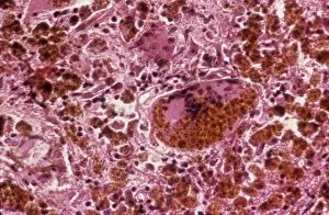
Excessive iron absorption. Light micrograph of a section through tissue showing haemochromatosis, a physiological disorder where excessive amounts of iron are absorbed by the body

Background imageHistopathological Collection: Gauchers disease, light micrograph

Gauchers disease, light micrograph
Gauchers disease. Light micrograph of a section through the spleen of a patient with Gauchers disease. This is an inherited metabolic disorder in which a fatty substance called glucocerebroside

Background imageHistopathological Collection: Glomerulosclerosis, TEM C016 / 5836

Glomerulosclerosis, TEM C016 / 5836
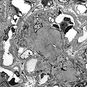
Glomerulosclerosis. Transmission electron micrograph (TEM) of a section through through the glomerulus of a kidney affected by glomerulosclerosis

Background imageHistopathological Collection: Alveolar soft part sarcoma, TEM C016 / 5826

Alveolar soft part sarcoma, TEM C016 / 5826
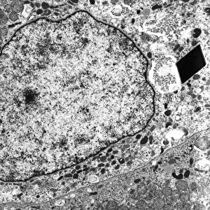
Alveolar soft part sarcoma. Transmission electron micrograph (TEM) of a section through an alveolar soft part sarcoma (ASPS) showing a characteristic rhomboid crystal (black, centre)

Background imageHistopathological Collection: Alveolar soft part sarcoma, TEM C016 / 5816

Alveolar soft part sarcoma, TEM C016 / 5816
Alveolar soft part sarcoma. Transmission electron micrograph (TEM) of a section through an alveolar soft part sarcoma (ASPS) showing a characteristic rhomboid crystal (black)

Background imageHistopathological Collection: Mitochondrial inclusions, TEM C016 / 5807

Mitochondrial inclusions, TEM C016 / 5807
Mitochondrial inclusions. Transmission electron micrograph (TEM) of a section through a mitochondrion showing crystalline inclusions (thick parallel rods) in the intermembrane spaces

Background imageHistopathological Collection: Mitochondrial inclusions, TEM C016 / 5806

Mitochondrial inclusions, TEM C016 / 5806
Mitochondrial inclusions. Transmission electron micrograph (TEM) of a section through a mitochondrion showing crystalline inclusions (thick parallel rods) in the intermembrane spaces

Background imageHistopathological Collection: Prostate cancer cell, SEM C013 / 5012

Prostate cancer cell, SEM C013 / 5012
Prostate cancer cell, coloured scanning electron micrograph (SEM). The uneven surface of this cell is typical of cancer cells

Background imageHistopathological Collection: Prostate cancer cell, SEM C013 / 5011

Prostate cancer cell, SEM C013 / 5011
Prostate cancer cell, coloured scanning electron micrograph (SEM). The uneven surface of this cell is typical of cancer cells

Background imageHistopathological Collection: Crystals in seminal fluid

Crystals in seminal fluid. Polarised light micrograph of a sample of abnormal seminal fluid. The sample contains crystals, which may indicate a problem with the prostate

Background imageHistopathological Collection: Psoriasis, light micrograph

Psoriasis, light micrograph
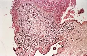
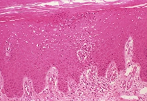
Psoriasis. Light micrograph of a section through a psoriasis pustule. Psoriasis is a chronic skin disease causing inflammation and scaly lesions

Background imageHistopathological Collection: Typhoid nodules, light micrograph

Typhoid nodules, light micrograph
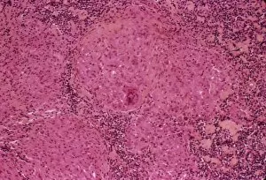
Typhoid nodules, coloured light micrograph. Section through a lymph node of a patient with typhoid, showing macrophages (large cells, pink) forming typhoid nodules

Background imageHistopathological Collection: Liver tuberculosis, light micrograph

Liver tuberculosis, light micrograph
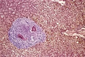
Liver tuberculosis. Coloured light micrograph of a section through the liver of a patient with miliary tuberculosis (TB). A tubercle, a nodular lesion of infected dead tissue, is seen at left

Background imageHistopathological Collection: Miliary tuberculosis, light micrograph

Miliary tuberculosis, light micrograph
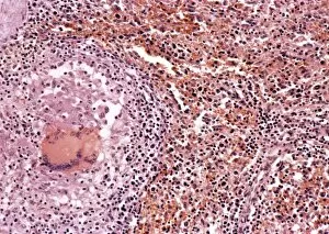
Miliary tuberculosis. Light micrograph of a section through a single miliary tubercle (left) in a patients lung. Tubercles are nodular lesions of infected dead tissue that arise from tuberculosis (TB)

Background imageHistopathological Collection: Systemic lupus erythematosus

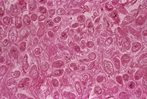
Systemic lupus erythematosus. Light micrograph of a section through a glomerulus (blood filtration structure) from kidney tissue in a case of SLE

Background imageHistopathological Collection: Skin inflammation, light micrograph

Skin inflammation, light micrograph
Skin inflammation. Light micrograph of a section through skin showing inflamed capillaries (lower left and upper right) caused by capillaritis, also known as pigmented purpurea

Background imageHistopathological Collection: Bilharzia infection, ureter tissue

Bilharzia infection, ureter tissue
Bilharzia infection. Light micrograph of human ureter tissue that contains numerous eggs from Schistosoma flukes. The ureters are tubes that carry urine from the kidneys to the bladder

Background imageHistopathological Collection: Skin pigmentation in Addisons disease

Skin pigmentation in Addisons disease
Skin pigmentation from Addisons disease. Light micrograph of a section through the skin of a patient with Addisons disease

Background imageHistopathological Collection: Marie-Francois Bichat, pathologist

Marie-Francois Bichat, pathologist
Marie-Francois Xavier Bichat (1771-1802), French pathologist and founder of modern histology. Bichat noted that various organs consist of several components, or tissues


All Professionally Made to Order for Quick Shipping

Histopathological investigations offer valuable insights into various diseases and conditions affecting the human body. From identifying specific cellular abnormalities to understanding the progression of life-threatening illnesses, histopathology plays a crucial role in diagnosis and treatment. Dohle bodies in blood cells, as seen through a micrograph, provide essential clues for medical professionals studying infections or inflammatory disorders. These distinctive blue-grey structures within neutrophils can indicate underlying health issues. In another micrograph, acute promyelocytic leukemia is evident with its characteristic abnormal promyelocytes. This aggressive form of leukemia requires prompt identification for effective management. A light micrograph showcasing ovarian cancer (C015 / 7103) highlights the importance of early detection in combating this deadly disease that affects women worldwide. Cystic fibrosis, a genetic disorder primarily affecting the respiratory system, can be identified through histopathological examinations revealing thick mucus accumulation and damaged lung tissue. Gout crystals are easily recognizable under microscopic examination due to their needle-like appearance. The presence of these crystals helps confirm gout as the cause behind painful joint inflammation. Examining lymphocytes within hair follicles using scanning electron microscopy provides researchers with valuable data about immune responses occurring at localized sites on our skin. Biopsy slides (F007 / 0316) play an integral role in diagnosing various conditions by allowing pathologists to analyze tissue samples under high magnification. These slides aid in determining appropriate treatment plans tailored to each patient's needs. Light micrographs depicting testicular cancer highlight distinct cellular changes that help differentiate it from other forms of malignancies originating from testicular tissues. Dementia, characterized by cognitive decline and memory loss, leaves its mark on brain tissue visible through light microscopy. Identifying key pathological features aids in understanding this complex neurodegenerative condition better. Multiple myeloma manifests itself within bone marrow plasma cells and can be observed via light microscopy techniques. Accurate diagnosis enables healthcare professionals to devise targeted treatment strategies.