
Imagery Collection (page 8)
"Unveiling the Power of Imagery: From His Masters Breath to Venera 13's Venus Surface" Step into a world where imagination knows no bounds


All Professionally Made to Order for Quick Shipping
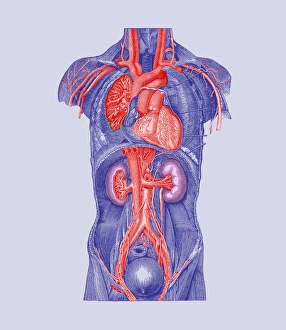
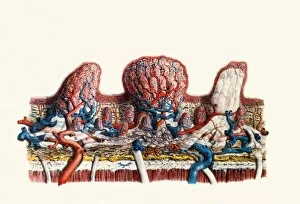
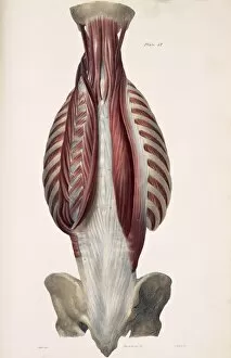
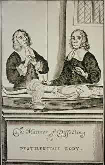
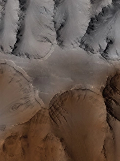
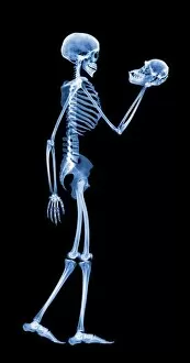
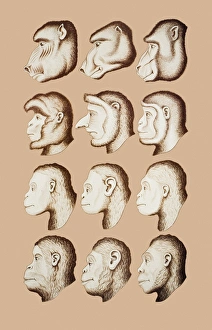
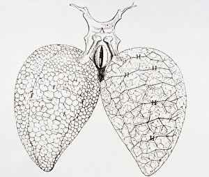
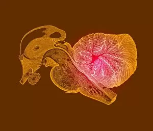
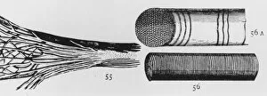
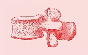
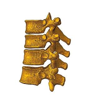
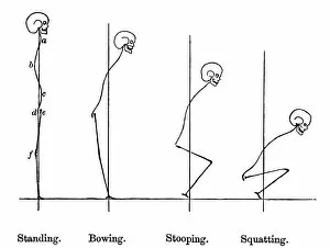
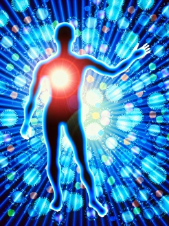
"Unveiling the Power of Imagery: From His Masters Breath to Venera 13's Venus Surface" Step into a world where imagination knows no bounds, as we embark on a journey through captivating imagery. Satire takes center stage with "His Masters Breath, " an artistic masterpiece that challenges societal norms and provokes thought. Behold the mesmerizing set of glass eyeballs, each one capturing stories untold and secrets hidden within their crystalline depths. Transport yourself to another planet with Venera 13's breathtaking photos, revealing the enigmatic surface of Venus in all its glory. Feel the adrenaline rush at Yugoslavia's Sokol Rally in Belgrade, where speed and precision merge into a symphony of motion captured forever in time. Leonardo da Vinci's iconic Vitruvian Man takes a twist as he bears a flare deep within his chest, symbolizing the eternal flame of passion burning inside us all. Delve into ancient wisdom with computer artwork depicting Chakra energy points, unlocking the mystical power that lies dormant within our souls. The vast expanse of Saudi Arabian desert stretches before you like an endless canvas; let your mind wander amidst its golden dunes. Explore Descartes' groundbreaking optics theory from the 17th century, unraveling how light shapes our perception and understanding of reality itself. Witness biomechanics come alive through historical artwork, showcasing intricate depictions of human movement and form throughout history. Marvel at the majestic Pyramids at Giza standing tall against time's relentless passage; these architectural wonders continue to captivate hearts worldwide. Discover serenity amidst chaos as Buddha's stone head rests peacefully in the roots of a Bodhi tree at Wat Mahathat in Ayutthaya - an image that embodies tranquility and enlightenment. Study every contour meticulously as you explore detailed illustrations highlighting muscles intricately woven within our necks - marvels crafted by nature herself.