
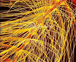
Insecta Collection (page 100)
"Insecta: A Kaleidoscope of Nature's Wonders" Ornithoptera alexandrae, commonly known as Queen Alexandra's birdwing butterfly


All Professionally Made to Order for Quick Shipping
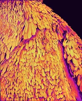
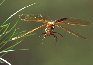
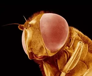
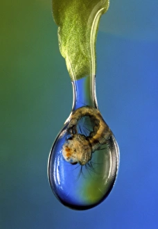
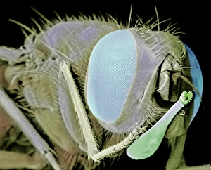
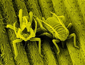
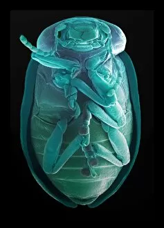
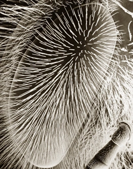
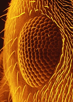
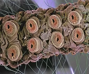
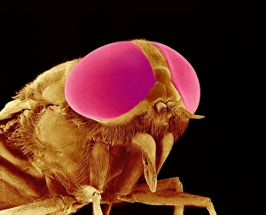
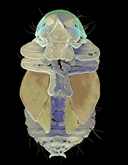
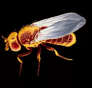
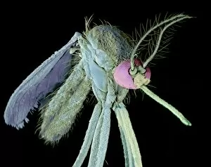
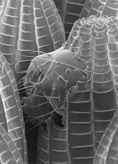
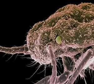
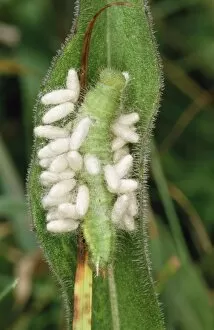
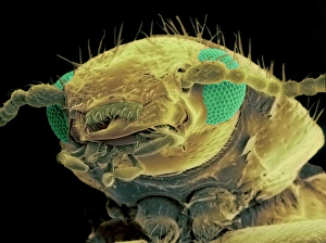
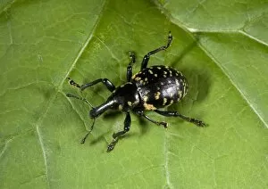
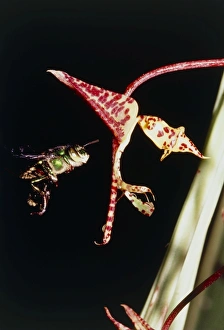
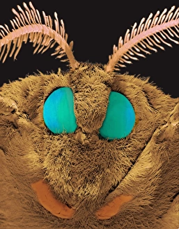
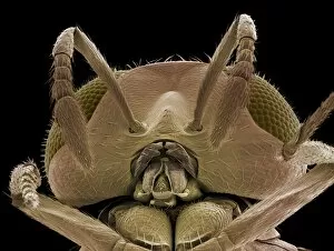
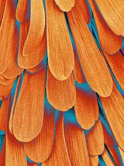
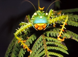
"Insecta: A Kaleidoscope of Nature's Wonders" Ornithoptera alexandrae, commonly known as Queen Alexandra's birdwing butterfly, showcases its majestic beauty with vibrant hues and intricate patterns. Honey bees diligently work on a honeycomb, their teamwork and precision creating the sweet nectar we all enjoy. Entomology specimens offer a glimpse into the incredible diversity of insects, each specimen telling a unique story of adaptation and survival. The Red Admiral (Vanessa atalanta) takes a moment to sunbathe on a plant, its wings spread wide to soak up the warm rays of sunlight. Common blue butterflies (Polyommatus icarus) gracefully bask in the morning light at Vealand Farm in Devon, UK, painting the landscape with delicate shades of blue. The Chrysina limbata silver chafer beetle shines like molten metal under sunlight, reminding us that even tiny creatures can possess extraordinary allure. Inachis io or peacock butterfly captivates with its iridescent wings resembling an artist's masterpiece as it flutters through meadows and gardens. The Red Admiral - Vanessa atalanta - embarks on a quest for nectar amidst blooming Common Boneset in Baden-Wurttemberg, Germany; nature's harmonious dance between insect and flower unfolds before our eyes. Sea green swallowtail butterfly glides effortlessly through coastal landscapes, enchanting observers with its ethereal beauty against azure skies and emerald waves. Under microscopic scrutiny lies the intricate world of fruit flies (SEM Z340 / 0768), revealing their astonishing complexity despite their diminutive size – yet another reminder that appearances can be deceiving. Phoebis sennae or cloudless sulphur butterfly brings sunshine wherever it goes; its golden wings brightening up gardens and meadows with their radiant glow.