Fraud Blocker Skip to main content

Jbu Colouring Collection

"Exploring the Vibrant World of JBU Colouring

Background imageJbu Colouring Collection: False col SEM of nematode worm on sample of peat

False col SEM of nematode worm on sample of peat
False-colour scanning electron micrograph of a nematode worm on a sample of peat. The micrograph shows the layer of " green slime" which often appears on peat & other organic surfaces

Background imageJbu Colouring Collection: False-colour SEM of a nematode worm & Acarus siro

False-colour SEM of a nematode worm & Acarus siro

Background imageJbu Colouring Collection: 2nd of the F / col SEMs of caterpillars hatching

2nd of the F / col SEMs of caterpillars hatching
Second of 5 false-colour scanning electron micro- graphs of caterpillars hatching from the eggs of the large white butterfly, Pieris brassicae

Background imageJbu Colouring Collection: SEM of caterpillars hatching from eggs

SEM of caterpillars hatching from eggs
Fourth of 5 false-colour scanning electron micro- graphs of caterpillars hatching from the eggs of the large white butterfly, Pieris brassicae

Background imageJbu Colouring Collection: 5th of the F / col SEMs of caterpillars hatching

5th of the F / col SEMs of caterpillars hatching
Fifth of 5 false-colour scanning electron microgr- aphs of caterpillars hatching from the eggs of the the large white butterfly, Pieris brassicae

Background imageJbu Colouring Collection: Fal-col TEM sequence of budding Aids virus

Fal-col TEM sequence of budding Aids virus

Background imageJbu Colouring Collection: Open stoma on tobacco leaf

Open stoma on tobacco leaf
False-colour scanning electron micrograph of an open stoma, or pore, on the surface of a leaf of the tobacco plant, Nicotiana tabacum

Background imageJbu Colouring Collection: Stoma on sepal of primula flower

Stoma on sepal of primula flower
False-colour scanning electron micrograph of a stoma, or pore, on a sepal of the primula flower, Primula malacoides. The stoma appears to be open, but is in fact closed

Background imageJbu Colouring Collection: SEM of pollen grains of Common Mallow

SEM of pollen grains of Common Mallow
False-colour scanning electron micrograph of pollen grains of Common Mallow, Malva sylvestris. The grains are spherical & about 108 micrometres across

Background imageJbu Colouring Collection: False-colour SEM of cocksfoot grass

False-colour SEM of cocksfoot grass
False-colour scanning electron micrograph (SEM) of two pollen grains of cocksfoot grass, Dactylis glomerata, a major cause of hay fever (allergic rhinitis)

Background imageJbu Colouring Collection: Cocksfoot pollen

Cocksfoot pollen
False-colour scanning electron micrograph (SEM) of two pollen grains of cocksfoot grass, Dactylis glomerata, a major cause of hay fever (allergic rhinitis)

Background imageJbu Colouring Collection: Pollen on honeybee face

Pollen on honeybee face
False-colour scanning electron micrograph of unidentified pollen grains on the hairs of the face of a worker honey bee, Apis mellifera

Background imageJbu Colouring Collection: Pollen of Common Mallow flower

Pollen of Common Mallow flower
False-colour scanning electron micrograph of pollen grains of Common Mallow, Malva sylvestris. The grains are spherical & about 108 micrometres across


All Professionally Made to Order for Quick Shipping

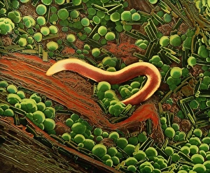
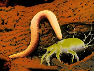
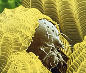
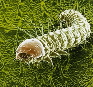
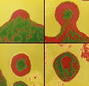
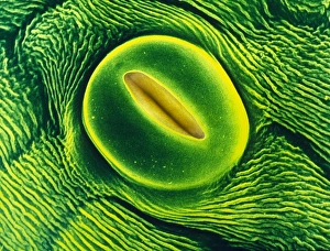
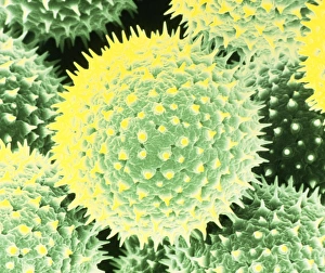
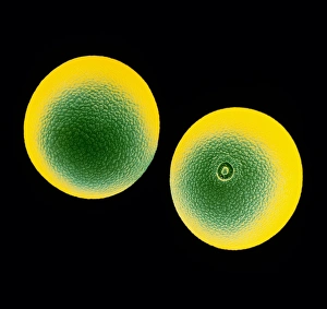
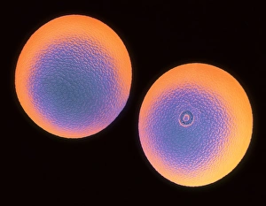
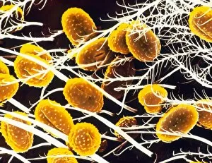
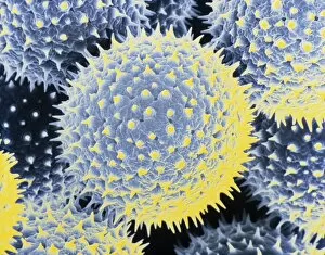
"Exploring the Vibrant World of JBU Colouring: From Nematode Worms to Pollen Grains" Dive into the mesmerizing world as we uncover stunning false-colour scanning electron microscope (SEM) images. Starting with a captivating SEM image, witness the intricate details of a nematode worm on a sample of peat, showcasing its unique structure and patterns. Moving forward, another breathtaking false-colour SEM reveals the fascinating encounter between a nematode worm and Acarus siro, offering an up-close look at their interaction in vivid detail. Shifting our focus to nature's wonders, feast your eyes on two remarkable SEMs capturing caterpillars hatching from eggs. The second image showcases these tiny creatures emerging into the world while highlighting their delicate features. Additionally, we present you with the fifth instalment of this series - yet another astonishing false-colour SEM displaying caterpillars hatching from their protective shells. Delving deeper into microscopic marvels, prepare to be amazed by a false-colour transmission electron microscope (TEM) sequence illustrating budding Aids virus particles. This extraordinary visual journey unveils the complex process through which these viruses replicate and spread. But it doesn't stop there. Witness bread mould like never before as we unveil an enchanting SEM image that exposes its intricate filaments and structures. Continuing our exploration through plant life, get ready for an awe-inspiring view of an open stoma on a tobacco leaf - revealing nature's ingenious design for gas exchange in plants. Furthermore, discover another hidden gem within primula flowers as we zoom in on a stoma located on its sepal. As we conclude this captivating journey through JBU colouring masterpieces, immerse yourself in the beauty of pollen grains from Common Mallow captured under high-resolution SEM imaging techniques - unveiling their diverse shapes and textures.