
Joint Disease Collection
"Exploring the World of Joint Disease: A Visual Journey" Osteoarthritis of the hip, as seen in this X-ray, reveals the wear and tear on the joint


All Professionally Made to Order for Quick Shipping
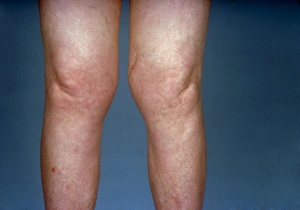
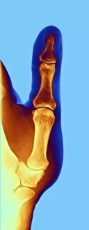
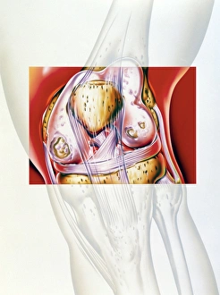
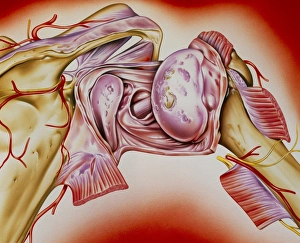
"Exploring the World of Joint Disease: A Visual Journey" Osteoarthritis of the hip, as seen in this X-ray, reveals the wear and tear on the joint, causing pain and stiffness. Arthrosis of the hand captured in X-ray F006/4616 showcases the degeneration of joints, affecting daily activities like gripping or writing. Take a glimpse at knees affected by osteoarthritis through this striking view, highlighting the impact on mobility and quality of life. Delving deeper into arthrosis of the hand with X-ray F006/4605, we witness how joint damage can hinder dexterity and finger movements. Another perspective on arthrosis is revealed in X-ray F006/4595, emphasizing its debilitating effects on hand functionality and comfort. The intricate details showcased in X-ray F006/4598 expose how arthrosis can distort joint structures within our hands over time. Artwork depicting arthritis of a thumb joint brings to life both its physical manifestation and its impact on everyday tasks involving grip or pinch motions. Witnessing an artwork showcasing rheumatoid arthritis of the knee reminds us that this autoimmune disease affects not only joints but also overall health and well-being. An artistic representation combining an arthritic knee with NSAID drug mechanism serves as a visual reminder that medical interventions are available to alleviate symptoms associated with joint diseases. This captivating artwork portrays rheumatoid arthritis affecting a shoulder joint—a poignant reminder that these conditions can manifest throughout various parts of our bodies beyond just hands or knees.