mail_outline sales@mediastorehouse.com
Framed Print
Canvas Print
Metal Print
Photographic Print
Poster Print
Fine Art Print
Jigsaw Puzzle
Photo Mug
Greetings Card
Cushion
Mouse Mat
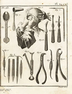
18th century lacrimal gland operation and surgical equipment18th century lacrimal gland operation 6, and dental surgical equipment: silver spoon to cover the eye 1, cauterizing funnel 2 and cauterizer 3, bone perforator 4, teeth scaler 5, probe 7, splints 8
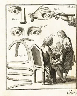
Eye surgery, 18th centuryLacrimal gland conditions 1, 2, bandages for lacrimal gland swelling 3, and cataract surgery 4, 5, 18th century. Copperplate engraving by Robert Benard from Denis Diderots Encyclopedia, Pellet
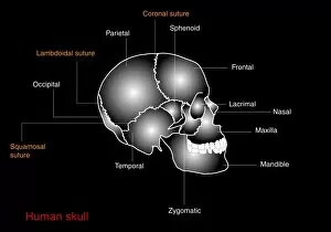
Human skull anatomy, diagramHuman skull anatomy. Diagram showing the structure and anatomy of a human skull, including the sutures (jagged lines), the joints between the fused skull bones
Lacrimal gland, light micrographLacrimal gland. Light micrograph of a section through a lacrimal gland. The lacrimal glands, which are situated one above each eye, secrete tears
Lacrimal gland, SEM
Human skull anatomy, artworkHuman skull anatomy. Artwork showing the structure and anatomy of a human skull, including the sutures (jagged lines), the joints between the fused skull bones
Skull bones, artworkSkull bones. Computer artwork showing the major bones of the human skull. These are: frontal (purple, upper left), parietal (green/beige, upper right), occipital (blue, lower right), temporal (pink)