
Lipid Collection
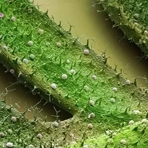
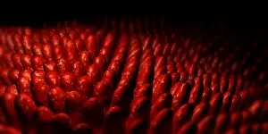
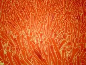
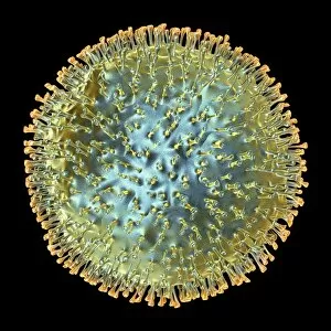
"Lipid: Unveiling the Intricate World of Fats and Cholesterol" Exploring the delicate beauty of nature, we observe the French lavender leaf surface under SEM


All Professionally Made to Order for Quick Shipping
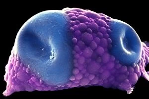
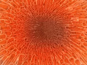
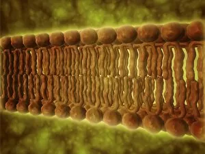
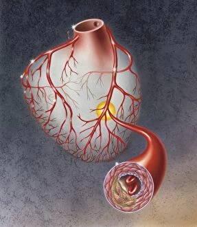
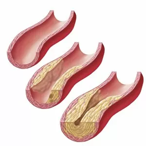
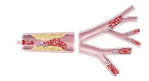
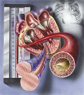
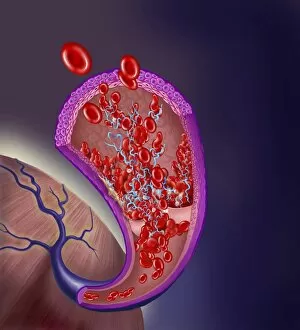
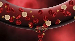
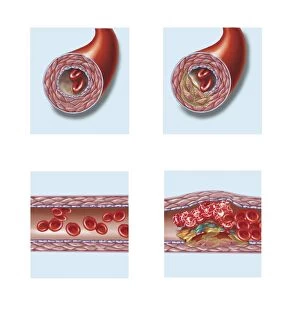
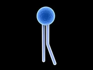
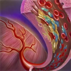
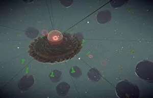
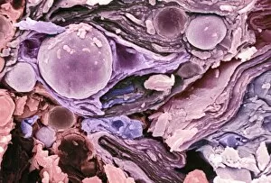
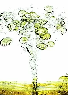
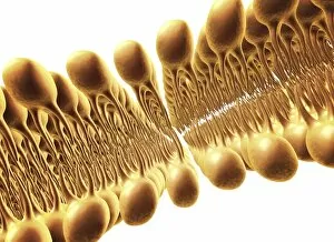
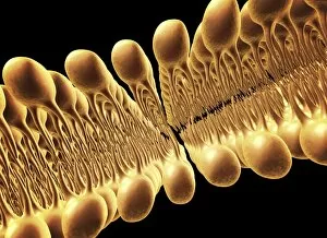
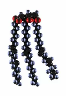
"Lipid: Unveiling the Intricate World of Fats and Cholesterol" Exploring the delicate beauty of nature, we observe the French lavender leaf surface under SEM, revealing intricate lipid structures. Delving deeper into our bodies, SEM showcases fat tissue with its unique lipid composition, providing essential energy storage and insulation. Returning to nature's wonders, SEM captures the mesmerizing details of the French lavender leaf surface once again, highlighting lipids' role in plant physiology. Art meets science as we visualize cell membrane lipid bilayers through artwork F007/1477, illustrating their crucial function in maintaining cellular integrity. Witnessing a distended fat cell under SEM (C013/5015), we comprehend how excessive lipid accumulation can lead to obesity-related health issues. Further magnifying a distended fat cell (SEM C013/5013), we gain insight into the consequences of unhealthy lifestyle choices on adipose tissue structure. Peering inside an artery with intestinal villi reveals a microscopic view that emphasizes lipids' involvement in nutrient absorption and transport within our bodies. In an intriguing display of cardiovascular health, one image portrays cholesterol buildup in an artery while another depicts plaque formation—a reminder of lipids' impact on heart disease development. Microscopic examination unveils phospholipids' elegant arrangement within cells—an architectural marvel critical for cellular functions such as signaling and barrier formation. A conceptual image beautifully illustrates intestinal villi—tiny finger-like projections enriched with lipids that enhance nutrient absorption efficiency within our digestive system. Examining arteries on a heart model exposes atherosclerotic plaque formation—an alarming consequence when excess lipids accumulate and obstruct blood flow throughout vital organs like the heart. Good or bad? The bloodstream harbors both types of cholesterol; this captivating image symbolizes their presence and highlights their contrasting effects on our overall health.