Fraud Blocker Skip to main content

Lipid Collection (#3)

"Lipid: Unveiling the Intricate World of Fats and Cholesterol" Exploring the delicate beauty of nature, we observe the French lavender leaf surface under SEM

Background imageLipid Collection: Avian influenza virus, TEM C016 / 5843

Avian influenza virus, TEM C016 / 5843
Avian influenza virus, type A strain H7N9, coloured transmission electron micrograph (TEM). This virus first emerged in the human population in China, in March 2013

Background imageLipid Collection: Avian influenza virus, TEM C016 / 5841

Avian influenza virus, TEM C016 / 5841
Avian influenza virus, type A strain H7N9, coloured transmission electron micrograph (TEM). This virus first emerged in the human population in China, in March 2013

Background imageLipid Collection: Avian influenza virus, TEM C016 / 5842

Avian influenza virus, TEM C016 / 5842
Avian influenza virus, type A strain H7N9, coloured transmission electron micrograph (TEM). This virus first emerged in the human population in China, in March 2013

Background imageLipid Collection: Avian influenza virus, TEM C016 / 2354

Avian influenza virus, TEM C016 / 2354
Avian influenza virus, type A strain H7N9, coloured transmission electron micrograph (TEM). This virus first emerged in the human population in China, in March 2013

Background imageLipid Collection: Avian influenza virus, TEM C016 / 2353

Avian influenza virus, TEM C016 / 2353
Avian influenza virus, type A strain H7N9, coloured transmission electron micrograph (TEM). This virus first emerged in the human population in China, in March 2013

Background imageLipid Collection: Avian influenza virus, TEM C016 / 2352

Avian influenza virus, TEM C016 / 2352
Avian influenza virus, type A strain H7N9, coloured transmission electron micrograph (TEM). This virus first emerged in the human population in China, in March 2013

Background imageLipid Collection: Avian influenza virus, TEM C016 / 2351

Avian influenza virus, TEM C016 / 2351
Avian influenza virus, type A strain H7N9, coloured transmission electron micrograph (TEM). This virus first emerged in the human population in China, in March 2013

Background imageLipid Collection: Global obesity pandemic, conceptual image C013 / 9664

Global obesity pandemic, conceptual image C013 / 9664
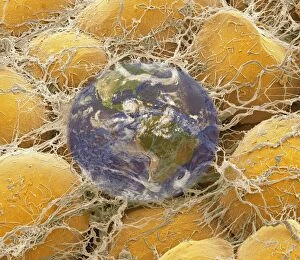
Global obesity pandemic, conceptual composite image. The background is a coloured scanning electron micrograph (SEM) of fat cells (adipocytes)

Background imageLipid Collection: Pore protein from staphylococcus aureus C013 / 9380

Pore protein from staphylococcus aureus C013 / 9380
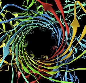
Structure of staphylococcal alpha-hemolysin, a heptameric transmembrane pore. Computer artwork of the ribbon structure of the pore forming-toxin from the bacteria Staphylococcus aureus

Background imageLipid Collection: Pore protein from staphylococcus aureus C013 / 9378

Pore protein from staphylococcus aureus C013 / 9378
Structure of staphylococcal alpha-hemolysin, a heptameric transmembrane pore. Computer artwork of the ribbon structure of the pore forming-toxin from the bacteria Staphylococcus aureus

Background imageLipid Collection: Pore protein from staphylococcus aureus C013 / 9379

Pore protein from staphylococcus aureus C013 / 9379
Structure of staphylococcal alpha-hemolysin, a heptameric transmembrane pore. Computer artwork of the ribbon structure of the pore forming-toxin from the bacteria Staphylococcus aureus

Background imageLipid Collection: Pore protein from staphylococcus aureus C013 / 9377

Pore protein from staphylococcus aureus C013 / 9377
Structure of staphylococcal alpha-hemolysin, a heptameric transmembrane pore. Computer artwork of the ribbon structure of the pore forming-toxin from the bacteria Staphylococcus aureus

Background imageLipid Collection: Cholesteryl ester transfer protein C013 / 8895

Cholesteryl ester transfer protein C013 / 8895
Cholesteryl ester transfer protein molecule. Computer model showing the structure of a molecule of cholesteryl ester transfer protein (CETP)

Background imageLipid Collection: Fat cells, SEM C013 / 5016

Fat cells, SEM C013 / 5016
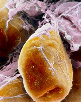
Fat cells. Coloured scanning electron micrograph (SEM) of several adipocytes (fat cells). Adipocytes form an insulating layer of fat in the body to store energy

Background imageLipid Collection: Distended fat cell, SEM C013 / 5014

Distended fat cell, SEM C013 / 5014
Distended fat cell. Coloured scanning electron micrograph (SEM) of a large adipocyte (fat cell, brown) distended by fat droplets (green)

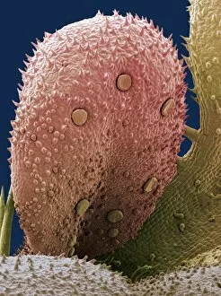
Background imageLipid Collection: Lavender leaf oil gland, SEM

Lavender leaf oil gland, SEM
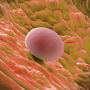
Lavender leaf oil gland. Coloured scanning electron micrograph (SEM) of an oil gland on the surface of a French lavender (Lavandula dentata) leaf

Background imageLipid Collection: Coloured SEM of fat cells in adipose tissue

Coloured SEM of fat cells in adipose tissue
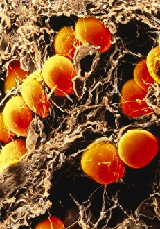
Fat cells. Coloured scanning electron micrograph of fat cells (orange), also known as adipocytes, in human adipose connective tissue

Background imageLipid Collection: False-colour SEM of a fat cell (adipocyte)

False-colour SEM of a fat cell (adipocyte)

Background imageLipid Collection: Fat cell

Fat cell. Computer artwork of a fat cell (adipocyte). Adipocytes form adipose tissue, which stores energy as an insulating layer of fat

Background imageLipid Collection: Adipose tissue, SEM

Adipose tissue, SEM
Adipose tissue. Coloured scanning electron micrograph (SEM) of adipose tissue. The tissue consists of adipocyte cells, whose lipid (fat) contents has been largely removed

Background imageLipid Collection: Influenzavirus A, TEM

Influenzavirus A, TEM
Influenzavirus A. Coloured transmission electron micrograph (TEM) of the influenza A virus, which can infect multiple species and is the most virulent human pathogen of the three influenza types

Background imageLipid Collection: Narrowed coronary artery

Narrowed coronary artery

Background imageLipid Collection: Atherosclerosis

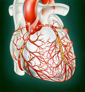
Atherosclerosis. Artwork of a human heart showing narrowing of the coronary arteries due to atherosclerosis, a build up of fatty deposits (atheroma, yellow) on the artery wall

Background imageLipid Collection: Narrowed artery, computer artwork

Narrowed artery, computer artwork
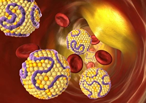
Narrowed artery. Computer artwork of a narrowed artery, showing red blood cells (red) and high levels of low density lipoprotein (LDL, yellow balls)

Background imageLipid Collection: Avian influenza virus, TEM

Avian influenza virus, TEM
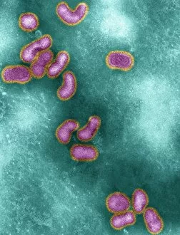
Avian influenza virus, type A strain H5N1, coloured transmission electron micrograph (TEM). This virus was isolated in Vietnam, during the avian flu outbreak in early 2004

Background imageLipid Collection: Algae growing in a photobioreactor

Algae growing in a photobioreactor with a researchers shadow. Algae are photosynthesising micro-organisms that can convert carbon dioxide into commercially valuable lipids

Background imageLipid Collection: Antibiotic cell membrane effect, artwork

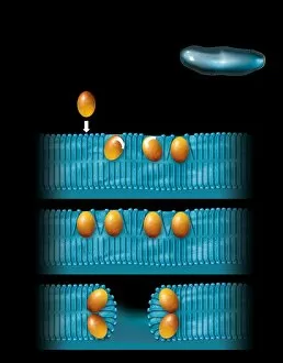
Antibiotic cell membrane effect, artwork
Antibiotic cell membrane effect. Artwork of the natural antibiotic peptide defensin (orange) disrupting the cell membrane of a bacterium (top right)

Background imageLipid Collection: Algae growing on petri dishes

Algae growing on petri dishes, Solazyme Inc. USA. Algae are photosynthesising micro-organisms. They convert carbon dioxide (CO2) into sugars, that are then metabolised into lipids

Background imageLipid Collection: Lipid bilayer membrane, molecular model

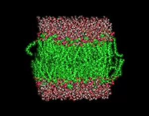
Lipid bilayer membrane, molecular model
Lipid bilayer membrane. Molecular model simulation of a lipid bilayer membrane consisting of POPC lipids (green), surrounded by water molecules (red and white)

Background imageLipid Collection: Algae research

Algae research. Researcher from Solazyme Inc. USA, examining a petri dish from an incubation room containing different algae strains. Algae are photosynthesising micro-organisms

Background imageLipid Collection: Liposomes, SEM

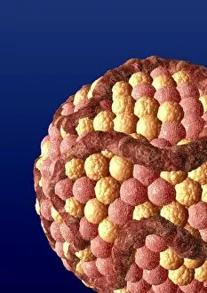
Liposomes, SEM

Background imageLipid Collection: Liposomes, TEM

Liposomes, TEM

Background imageLipid Collection: Algae fermentation bioreactor

Algae fermentation bioreactor, Solazyme Inc. USA. Algae are photosynthesising micro- organisms that can convert carbon dioxide into sugars

Background imageLipid Collection: Researcher with algae fermenter

Researcher with algae fermenter
Researcher with algae fermentation bioreactor, Solazyme Inc. USA. Algae are photosynthesising micro-organisms that can convert carbon dioxide (CO2) into sugars

Background imageLipid Collection: Fat cell, TEM

Fat cell, TEM
Fat cell. Coloured Coloured transmission electron micrograph (TEM) of a section through fat (adipose) tissue. Fat cells, or adipocytes

Background imageLipid Collection: Fat tissue, SEM

Fat tissue, SEM
Fat tissue. Coloured scanning electron micrograph (SEM) of fat cells (adipocytes, round) surrounded by fine strands of supportive connective tissue

Background imageLipid Collection: High cholesterol levels

High cholesterol levels. Computer artwork of balls of cholesterol. When there are high levels of low density lipoprotein (LDL)

Background imageLipid Collection: High cholesterol levels, artwork

High cholesterol levels, artwork
High cholesterol levels. Computer artwork balls of cholesterol in a blood vessel. Cholesterol is a lipid, a fat-like substance

Background imageLipid Collection: Cholesterol, artwork

Cholesterol, artwork

Background imageLipid Collection: Fatty acid synthase in complex with NADP+

Fatty acid synthase in complex with NADP+. Computer model showing the molecular structure of fatty acid synthase (FAS) complexed with nicotinamide adenine dinucleotide phosphate (NADP+)

Background imageLipid Collection: Milk-producing cell, artwork

Milk-producing cell, artwork
Milk-producing cell. Artwork of the anatomical structure of a lacteal (milk-producing) cell. Normal cell organelles include the nucleus (black, centre), the golgi apparatus (white)

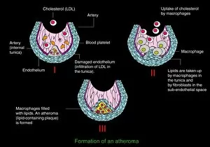
Background imageLipid Collection: Atheroma formation, diagram

Atheroma formation, diagram
Atheroma formation. Diagram showing the stages in the formation of an atheroma, a fatty plaque that forms on the inners walls of blood vessels in cardiovascular disease

Background imageLipid Collection: Milk-producing cell, diagram

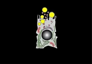
Milk-producing cell, diagram
Milk-producing cell. Diagram of the anatomical structure of a lacteal (milk-producing) cell. Normal cell organelles include the nucleus (black, centre), the golgi apparatus (white)

Background imageLipid Collection: Atheroma formation, artwork

Atheroma formation, artwork
Atheroma formation. Artwork showing the stages in the formation of an atheroma, a fatty plaque that forms on the inners walls of blood vessels in cardiovascular disease

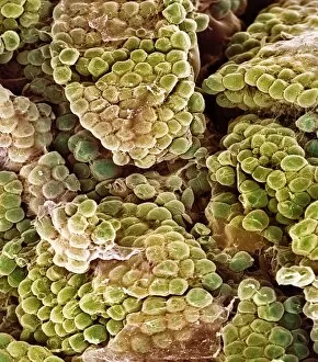
Background imageLipid Collection: Herb oil glands, SEM

Herb oil glands, SEM
Herb oil glands. Coloured scanning electron micrograph (SEM) of the underside of a thyme (Thymus vulgaris) leaf, showing the modified trichomes (hair cells, round)

Background imageLipid Collection: H1N1 Influenza A virus particles, TEM

H1N1 Influenza A virus particles, TEM
H1N1 Influenza A virus particles, coloured transmission electron micrograph (TEM). This influenza virus can infect multiple species

Background imageLipid Collection: Fat cells, TEM

Fat cells, TEM
Fat cells. Coloured transmission electron micrograph (TEM) of a section through fat (adipose) tissue. Each fat cell or adipocyte

Background imageLipid Collection: Fat cells, SEM

Fat cells, SEM
Fat cells, or adipocytes, coloured scanning electron micrograph (SEM). Adipocytes store energy as an insulating layer of fat and the majority of the cells volume is taken up by a large lipid droplet


All Professionally Made to Order for Quick Shipping

"Lipid: Unveiling the Intricate World of Fats and Cholesterol" Exploring the delicate beauty of nature, we observe the French lavender leaf surface under SEM, revealing intricate lipid structures. Delving deeper into our bodies, SEM showcases fat tissue with its unique lipid composition, providing essential energy storage and insulation. Returning to nature's wonders, SEM captures the mesmerizing details of the French lavender leaf surface once again, highlighting lipids' role in plant physiology. Art meets science as we visualize cell membrane lipid bilayers through artwork F007/1477, illustrating their crucial function in maintaining cellular integrity. Witnessing a distended fat cell under SEM (C013/5015), we comprehend how excessive lipid accumulation can lead to obesity-related health issues. Further magnifying a distended fat cell (SEM C013/5013), we gain insight into the consequences of unhealthy lifestyle choices on adipose tissue structure. Peering inside an artery with intestinal villi reveals a microscopic view that emphasizes lipids' involvement in nutrient absorption and transport within our bodies. In an intriguing display of cardiovascular health, one image portrays cholesterol buildup in an artery while another depicts plaque formation—a reminder of lipids' impact on heart disease development. Microscopic examination unveils phospholipids' elegant arrangement within cells—an architectural marvel critical for cellular functions such as signaling and barrier formation. A conceptual image beautifully illustrates intestinal villi—tiny finger-like projections enriched with lipids that enhance nutrient absorption efficiency within our digestive system. Examining arteries on a heart model exposes atherosclerotic plaque formation—an alarming consequence when excess lipids accumulate and obstruct blood flow throughout vital organs like the heart. Good or bad? The bloodstream harbors both types of cholesterol; this captivating image symbolizes their presence and highlights their contrasting effects on our overall health.