Fraud Blocker Skip to main content

Magnification Collection (page 26)

Magnification: Revealing the Hidden World From the intricate patterns of a snowflake to the delicate structure of a human cell

Background imageMagnification Collection: Linezolid antibiotic molecule

Linezolid antibiotic molecule. This synthetic antibiotic is used to treat infections that have become resistant to other antibiotics

Background imageMagnification Collection: Phentermine obesity drug molecule

Phentermine obesity drug molecule
Phentermine obesity drug, molecular model. This drug suppresses appetite. Atoms are represented as spheres and are colour-coded: hydrogen (white), carbon (grey) and nitrogen (blue)

Background imageMagnification Collection: Scanning tunnelling microscope, artwork F005 / 0813

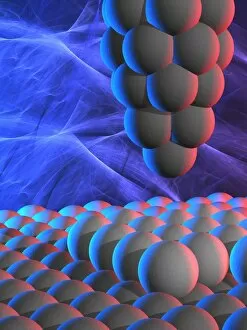
Scanning tunnelling microscope, artwork F005 / 0813
Scanning tunnelling microscope (STM), computer artwork. The STM uses a fine needle with an electric potential running through it to scan the surface of an object

Background imageMagnification Collection: Graves disease, light micrograph

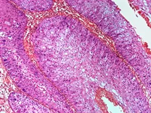
Graves disease, light micrograph
Graves disease. Light micrograph of a section through follicle from a thyroid gland affected by Graves disease. This is an autoimmune disease, where the thyroid is overactive

Background imageMagnification Collection: Blood clot, light micrograph

Blood clot, light micrograph
Blood clot. Light micrograph of a section through an arterial thrombus. Blood clots can form in an intact vessel that contains fatty deposits or one that is inflamed

Background imageMagnification Collection: Appendicitis, light micrograph

Appendicitis, light micrograph
Appendicitis. Light micrograph of a section through an inflamed appendix. There is an accumulation of white blood cells (dark pink) at bottom left
Background imageMagnification Collection: Wart, light micrograph

Wart, light micrograph
Wart. Light micrograph of a section through a wart (verruca vulgaris). The epidermis (purple) is irregularly thickened. Magnification: x200 when printed at 10 centimetres wide

Background imageMagnification Collection: Kidney disease, light micrograph

Kidney disease, light micrograph
Kidney disease. Light micrograph of a section through an inflamed kidney showing a glomerulus (coiled capillaries, purple). It is surrounded by vascular exudate and necrotic tubules

Background imageMagnification Collection: Spinal bone, light micrograph

Spinal bone, light micrograph
Spinal bone. Light micrograph of a section through developing bone from the vertebral column. Calcified bone is blue and uncalcified bone is red. Bone marrow is pink

Background imageMagnification Collection: Blood vessel, TEM

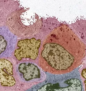
Blood vessel, TEM
Blood vessel. Coloured transmission electron micrograph (TEM) of a section through a blood vessel, showing platelets (blue) and red blood cells (red)

Background imageMagnification Collection: Corpora amylacea, light micrograph

Corpora amylacea, light micrograph
Corpora amylacea. Light micrograph of a section through the choroid plexus of brain, showing two corpora amylacea (purple, bottom). These small masses are a normal find in ageing brains

Background imageMagnification Collection: Fractured tendon, SEM F006 / 8643

Fractured tendon, SEM F006 / 8643
Fractured tendon, SEM

Background imageMagnification Collection: Fractured tendon, SEM F006 / 8642

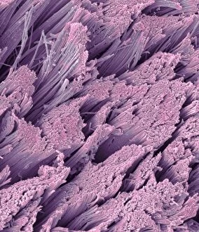
Fractured tendon, SEM F006 / 8642
Fractured tendon. Coloured scanning electron micrograph (SEM) of a fractured flexor tendon of the finger showing the collagen fibres

Background imageMagnification Collection: Lung cancer cells, SEM F006 / 8641

Lung cancer cells, SEM F006 / 8641
Lung cancer cells, coloured scanning electron micrograph (SEM). Magnification: x12, 000 when printed at 10 centimetres wide

Background imageMagnification Collection: Bone cancer cell, SEM F006 / 8639

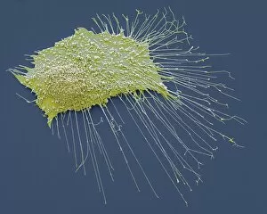
Bone cancer cell, SEM F006 / 8639
Bone cancer cell. Coloured scanning electron micrograph of an osteosarcoma cancer cell. Osteosarcoma is an aggressive malignant neoplasm arising from primitive transformed mesenchymal cells

Background imageMagnification Collection: Lung cancer cells, SEM F006 / 8640

Lung cancer cells, SEM F006 / 8640
Lung cancer cells, coloured scanning electron micrograph (SEM). Magnification: x12, 000 when printed at 10 centimetres wide

Background imageMagnification Collection: Bone cancer cell, SEM F006 / 8638

Bone cancer cell, SEM F006 / 8638
Bone cancer cell. Coloured scanning electron micrograph of an osteosarcoma cancer cell. Osteosarcoma is an aggressive malignant neoplasm arising from primitive transformed mesenchymal cells

Background imageMagnification Collection: Breast cancer, TEM F006 / 8633

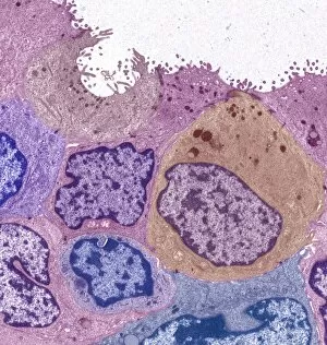
Breast cancer, TEM F006 / 8633
Breast cancer. Coloured transmission electron micrograph (TEM) of a section through a ductal carcinoma (cancer) of the breast showing malignant epithelial cells

Background imageMagnification Collection: Activated macrophage, SEM F006 / 8635

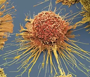
Activated macrophage, SEM F006 / 8635
Activated human macrophage, coloured scanning electron micrograph (SEM). Magnification: x2, 700 when printed at 10 centimetres wide

Background imageMagnification Collection: Activated macrophage, SEM F006 / 8634

Activated macrophage, SEM F006 / 8634
Activated human macrophage, coloured scanning electron micrograph (SEM). Magnification: x2, 700 when printed at 10 centimetres wide

Background imageMagnification Collection: Breast cancer, TEM F006 / 8632

Breast cancer, TEM F006 / 8632
Breast cancer. Coloured transmission electron micrograph (TEM) of a section through a ductal carcinoma (cancer) of the breast showing malignant epithelial cells

Background imageMagnification Collection: Data Analysis computer artwork C016 / 9197

Data Analysis computer artwork C016 / 9197
Analysis of data is a process of inspecting, cleaning, transforming, and modeling data with the goal of discovering useful information, suggesting conclusions, and supporting decision making

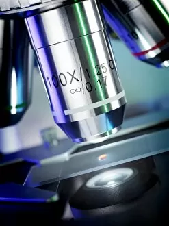
Background imageMagnification Collection: Light microscopy C016 / 9700

Light microscopy C016 / 9700
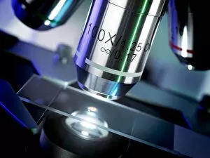
Light microscopy. Light microscope being used to magnify a blood sample

Background imageMagnification Collection: Light microscopy C016 / 9703

Light microscopy C016 / 9703
Light microscopy. Light microscope being used to magnify a blood sample

Background imageMagnification Collection: Data Analysis computer artwork C016 / 9187

Data Analysis computer artwork C016 / 9187
Analysis of data is a process of inspecting, cleaning, transforming, and modeling data with the goal of discovering useful information, suggesting conclusions, and supporting decision making

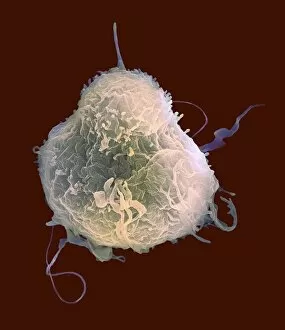
Background imageMagnification Collection: Lymphocyte white blood cell, SEM C016 / 9415

Lymphocyte white blood cell, SEM C016 / 9415
Lymphocyte white blood cell. Coloured scanning electron micrograph (SEM) of a lymphocyte white blood cell. Lymphocytes are involved in the immune systems defence mechanisms, lymph system

Background imageMagnification Collection: Data Analysis computer artwork C016 / 9196

Data Analysis computer artwork C016 / 9196
Analysis of data is a process of inspecting, cleaning, transforming, and modeling data with the goal of discovering useful information, suggesting conclusions, and supporting decision making

Background imageMagnification Collection: Light microscopy C016 / 9702

Light microscopy C016 / 9702
Light microscopy. Light microscope being used to magnify a blood sample

Background imageMagnification Collection: Data Analysis computer artwork C016 / 9202

Data Analysis computer artwork C016 / 9202
Conversion of numeric data into digital data. Analysis of data is a process of inspecting, cleaning, transforming, and modeling data with the goal of discovering useful information

Background imageMagnification Collection: Light microscopy C016 / 9701

Light microscopy C016 / 9701
Light microscopy. Light microscope being used to magnify a blood sample

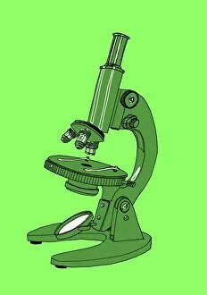
Background imageMagnification Collection: Light microscope, illustration C018 / 0737

Light microscope, illustration C018 / 0737
Light microscope. Illustration of a light microscope with three lenses and a mirror for reflecting light towards the object being viewed

Background imageMagnification Collection: Light microscope, illustration C018 / 0738

Light microscope, illustration C018 / 0738
Light microscope. Illustration of a light microscope with three lenses and a mirror for reflecting light towards the object being viewed

Background imageMagnification Collection: Comet dust grain model

Comet dust grain model (20, 000x magnification)

Background imageMagnification Collection: Bacillus sp. bacteria

Bacillus sp. bacteria
Bacteria are the most diverse and ubiquitous soil organisms present on Earth

Background imageMagnification Collection: Schem VIII from Robert Hookes Micrographia

Schem VIII from Robert Hookes Micrographia
Schem VIII located between pages 88 & 89 in Micrographia: or Some physical descriptions of minute bodies made by magnifying glasses, with observations and enquiries thereupon

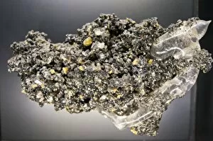
Background imageMagnification Collection: Malachite

Malachite comprises of (copper carbonate hydroxide). Malachite has distinctive green banding and belongs to the carbonate class

Background imageMagnification Collection: Difflugia Corona

Difflugia Corona
Freshwater Testate Amoebae. Magnification x 450

Background imageMagnification Collection: Lycopod

Lycopod

Background imageMagnification Collection: Neanura ?muscorum, plant mould

Neanura ?muscorum, plant mould
A magnifiied image of oak leaf mould. Specimen originates from Horstead, Norwich

Background imageMagnification Collection: Rusty screw

Rusty screw

Background imageMagnification Collection: Surface of a rusty screw

Surface of a rusty screw

Background imageMagnification Collection: Matchstick

Matchstick
Scanning electron microscope (SEM) image showing the fractured surface of a matchstick (x 400 on a standard 9 cm wide print)

Background imageMagnification Collection: Hydra sp

Hydra sp
Scanning electron microscope (SEM) image showing the stinging tentacles and mouth of the coelenterate Hydra (x 36 on a standard 9cm wide print)

Background imageMagnification Collection: Small fly, species unknown

Small fly, species unknown
Scanning electron microscope (SEM) of a fly head. Image displayed on the glass screens in the Darwin Centre, at the Natural History Museum, London

Background imageMagnification Collection: Woodlouse antenna

Woodlouse antenna
Scanning Electron Microscope (SEM) image of woodlouse antenna

Background imageMagnification Collection: Oats

Oats
A scanning electron microscope (SEM) image of oats, artificially coloured by computer

Background imageMagnification Collection: Hystrichopsylla talpae talpae, mole flea

Hystrichopsylla talpae talpae, mole flea
A macro photograph of the largest flea in the UK, the mole flea (Hystrichopsylla talpae talpae), which is common on small mammals throughout the UK


All Professionally Made to Order for Quick Shipping

Magnification: Revealing the Hidden World From the intricate patterns of a snowflake to the delicate structure of a human cell, magnification allows us to explore and unravel the mysteries of our world. Peering through the lens, we uncover secrets that would otherwise remain unseen. Take for instance Crysotile asbestos, its fibrous nature exposed under intense magnification, reminding us of its hidden dangers lurking within. In medical illustrations, an appendix with appendicitis comes alive as we zoom in closer. The inflamed tissues become vividly apparent, highlighting the urgency for surgical intervention. Moving deeper into our bodies, we discover the liver - a complex organ responsible for countless vital functions. Under magnification, its intricate network of cells and blood vessels reveals itself like an elaborate cityscape bustling with activity. But it's not just within ourselves that magnification unveils wonders; it extends beyond to nature's realm. A dandelion's fruiting head transforms into a mesmerizing display when observed up close – each tiny seed ready to embark on its own journey through wind and time. Fractals captivate our imagination as well – from Julia fractals with their infinite complexity to Mandelbrot fractals' self-repeating patterns. These mathematical marvels remind us of nature's innate ability to create beauty even in abstract forms. Anton van Leeuwenhoek revolutionized science by observing animalcules through his microscope in 1795. His discoveries opened up new frontiers in microbiology and forever changed our understanding of life at microscopic levels. Even seemingly ordinary creatures like snails hold surprises when examined closely; their teeth reveal intricate structures designed for efficient feeding and survival. Ponds teem with life too - water fleas gracefully glide alongside green algae like Volvox aureus. Magnified images transport us into this miniature ecosystem where every organism plays a crucial role in maintaining balance and harmony. And finally, we come full circle with snowflakes.