
Magnification Collection (page 3)
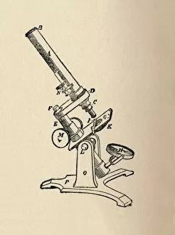
Magnification: Revealing the Hidden World From the intricate patterns of a snowflake to the delicate structure of a human cell


All Professionally Made to Order for Quick Shipping
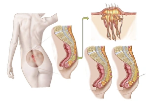
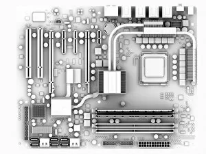
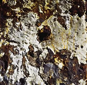
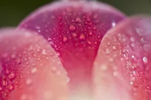
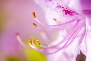
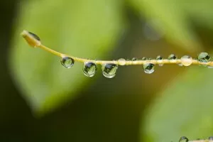
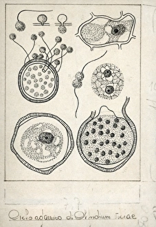
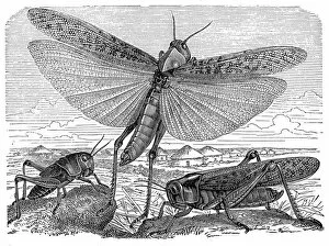
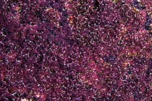
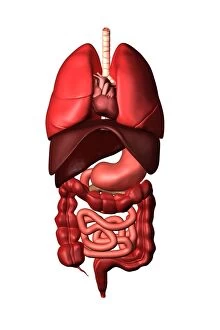
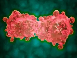
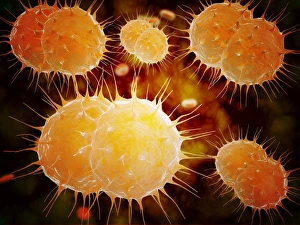
Magnification: Revealing the Hidden World From the intricate patterns of a snowflake to the delicate structure of a human cell, magnification allows us to explore and unravel the mysteries of our world. Peering through the lens, we uncover secrets that would otherwise remain unseen. Take for instance Crysotile asbestos, its fibrous nature exposed under intense magnification, reminding us of its hidden dangers lurking within. In medical illustrations, an appendix with appendicitis comes alive as we zoom in closer. The inflamed tissues become vividly apparent, highlighting the urgency for surgical intervention. Moving deeper into our bodies, we discover the liver - a complex organ responsible for countless vital functions. Under magnification, its intricate network of cells and blood vessels reveals itself like an elaborate cityscape bustling with activity. But it's not just within ourselves that magnification unveils wonders; it extends beyond to nature's realm. A dandelion's fruiting head transforms into a mesmerizing display when observed up close – each tiny seed ready to embark on its own journey through wind and time. Fractals captivate our imagination as well – from Julia fractals with their infinite complexity to Mandelbrot fractals' self-repeating patterns. These mathematical marvels remind us of nature's innate ability to create beauty even in abstract forms. Anton van Leeuwenhoek revolutionized science by observing animalcules through his microscope in 1795. His discoveries opened up new frontiers in microbiology and forever changed our understanding of life at microscopic levels. Even seemingly ordinary creatures like snails hold surprises when examined closely; their teeth reveal intricate structures designed for efficient feeding and survival. Ponds teem with life too - water fleas gracefully glide alongside green algae like Volvox aureus. Magnified images transport us into this miniature ecosystem where every organism plays a crucial role in maintaining balance and harmony. And finally, we come full circle with snowflakes.