Fraud Blocker Skip to main content

Magnification Collection (page 7)

Magnification: Revealing the Hidden World From the intricate patterns of a snowflake to the delicate structure of a human cell

Background imageMagnification Collection: Detail of a large coral polyp in Komodo National Park, Indonesia

Detail of a large coral polyp in Komodo National Park, Indonesia. This tropical region in Indonesia is known for its spectacular coral reefs and high marine biodiversity

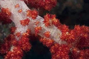
Background imageMagnification Collection: Red tree coral on a Fijian reef

Red tree coral on a Fijian reef

Background imageMagnification Collection: Side view of a pale cream colored thorny seahorse

Side view of a pale cream colored thorny seahorse (Hippocampus histrix) against a dark background, Komodo National Park, Indonesia

Background imageMagnification Collection: A pair of mushroom coral shrimp on pink mushroom coral

A pair of mushroom coral shrimp on pink mushroom coral
A pair of mushroom coral shrimp (Periclimenes Kororensis) on pink mushroom coral, Komodo National Park, Indonesia

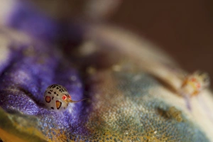
Background imageMagnification Collection: Close-up view of a ladybug amphipod, Cyproidea species

Close-up view of a ladybug amphipod, Cyproidea species
Ladybug amphipod, Cyproidea species, side view, taken at Cannibal Rock, Komodo National Park, Indonesia

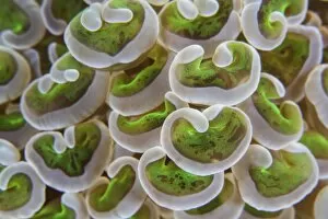
Background imageMagnification Collection: Detail of anchor coral tentacles growing on a reef in Indonesia

Detail of anchor coral tentacles growing on a reef in Indonesia
Detail of anchor coral tentacles (Euphyllia sp.) growing on a reef in Indonesia. This area is known for its high marine biodiversity

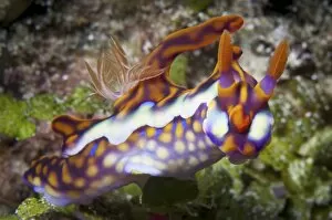
Background imageMagnification Collection: Yellow-Ridged Ceratosoma nudibranch

Yellow-Ridged Ceratosoma nudibranch
Yellow-Ridged Ceratosoma (Ceratosoma flavicostatum) nudibranch, head on view, Komodo National Park, Indonesia

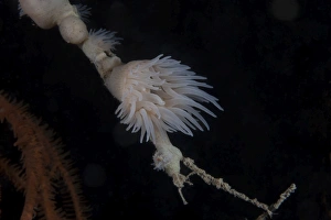
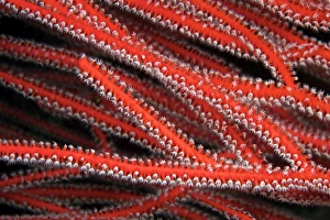
Background imageMagnification Collection: Gorgonain fan with polyps out feeding, Papua New Guinea

Gorgonain fan with polyps out feeding, Papua New Guinea

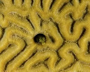
Background imageMagnification Collection: Small blenny in brain coral, Curacao

Small blenny in brain coral, Curacao

Background imageMagnification Collection: A pair of Risbecia tryoni nudibranch, Beqa Lagoon, Fiji

A pair of Risbecia tryoni nudibranch, Beqa Lagoon, Fiji

Background imageMagnification Collection: Chromodoris kuniei nudibranch, Beqa Lagoon, Fiji

Chromodoris kuniei nudibranch, Beqa Lagoon, Fiji

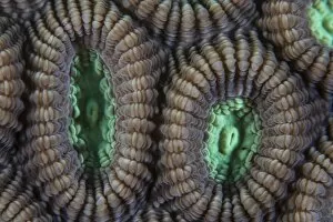
Background imageMagnification Collection: Detail of the polyps of a reef-building coral

Detail of the polyps of a reef-building coral growing in Indonesia. This beautiful, tropical region is home to an incredible variety of marine life

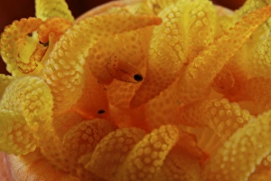
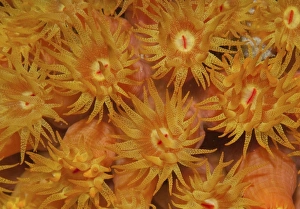
Background imageMagnification Collection: Detailed view of yellow tube coral (Tubastrea) polyps with parasites

Detailed view of yellow tube coral (Tubastrea) polyps with parasites, Bonaire, Caribbean Netherlands

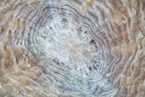
Background imageMagnification Collection: Abstract of a mushroom coral growing on a reef in Indonesia

Abstract of a mushroom coral growing on a reef in Indonesia
Abstract of a mushroom coral (Fungia sp.) growing on a reef in Indonesia. This tropical region, within the Coral Triangle, is home to an incredible variety of marine life

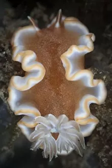
Background imageMagnification Collection: A nudibranch crawls across the seafloor

A nudibranch crawls across the seafloor
A nudibranch (Glossodoris rufomarginatus) crawls across the seafloor of Lembeh Strait, Indonesia

Background imageMagnification Collection: Small hermit crab living in an abandoned coral polyp hole, Fiji

Small hermit crab living in an abandoned coral polyp hole, Fiji

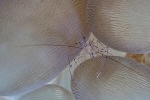
Background imageMagnification Collection: A saltwater shrimp living on bubble coral in Lembeh Strait

A saltwater shrimp living on bubble coral in Lembeh Strait
An almost invisible shrimp (Vir philippinensis) lives among the bulbous tentacles of a bubble coral on a reef in Lembeh Strait, Indonesia. This area is known for its high marine biodiversity

Background imageMagnification Collection: Cup coral polyps hang under a ledge on a reef in Fiji

Cup coral polyps hang under a ledge on a reef in Fiji

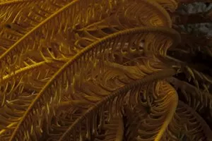
Background imageMagnification Collection: Close-up image of a yellow crinoid on a Fijian reef

Close-up image of a yellow crinoid on a Fijian reef

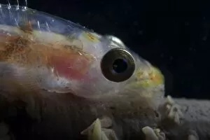
Background imageMagnification Collection: Close-up of a whip coral goby, Beqa Lagoon, Fiji

Close-up of a whip coral goby, Beqa Lagoon, Fiji

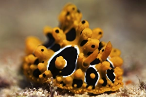
Background imageMagnification Collection: A brightly colored nudibranch feeds on algae, Fiji

A brightly colored nudibranch feeds on algae, Fiji

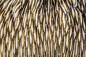
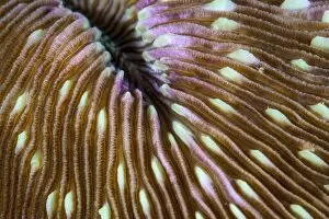
Background imageMagnification Collection: Detail of a mushroom coral growing on a reef in Indonesia

Detail of a mushroom coral growing on a reef in Indonesia
Detail of a mushroom coral (Fungia sp.) growing on a reef in Indonesia. This tropical region, within the Coral Triangle, is home to an incredible variety of marine life

Background imageMagnification Collection: Detail of a pin cushion starfish on a reef in Indonesia

Detail of a pin cushion starfish on a reef in Indonesia
Detail of a pin cushion starfish (Culcita sp.) on a reef in Indonesia

Background imageMagnification Collection: Detail of coral polyps on a reef in Lembeh Strait

Detail of coral polyps on a reef in Lembeh Strait
Detail of coral polyps (Diploastrea sp.) on a reef in Lembeh Strait, Indonesia

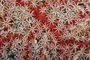
Background imageMagnification Collection: Detailed close-up view of soft coral polyps feeding, Fiji

Detailed close-up view of soft coral polyps feeding, Fiji

Background imageMagnification Collection: Close-up of orange cup coral in the Caribbean

Close-up of orange cup coral in the Caribbean

Background imageMagnification Collection: H-alpha Sun with active area

H-alpha Sun with active area

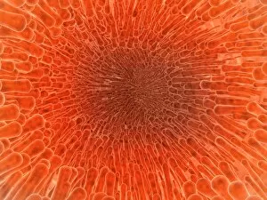
Background imageMagnification Collection: Microscopic view inside of the artery with intestinal villi

Microscopic view inside of the artery with intestinal villi

Background imageMagnification Collection: Conceptual image of synapse of neuron inside brain

Conceptual image of synapse of neuron inside brain

Background imageMagnification Collection: Heart with arteries showing cholesterol in one and plaque in the other

Heart with arteries showing cholesterol in one and plaque in the other
Heart with arteries showing cholesterol in one artery and atherosclerotic plaque in the other

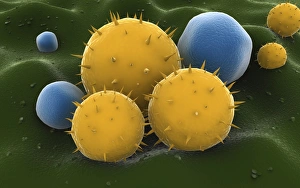
Background imageMagnification Collection: Microscopic visualization of grass pollen grains

Microscopic visualization of grass pollen grains

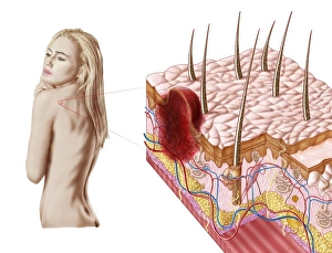
Background imageMagnification Collection: Illustration of an atypical growth on the skin that could be a sign of skin cancer

Illustration of an atypical growth on the skin that could be a sign of skin cancer
Illustration of an atypical growth on the skin that could be a warning sign of skin cancer. Sudden change in the appearance, shape, color, size

Background imageMagnification Collection: X-ray image of internal organs with large intestine highlighted in red

X-ray image of internal organs with large intestine highlighted in red

Background imageMagnification Collection: Microscopic view of cell

Microscopic view of cell

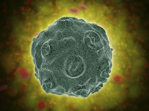
Background imageMagnification Collection: Single virus particle

Single virus particle

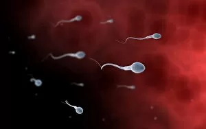
Background imageMagnification Collection: Conceptual image of sperm inside fallopian tube

Conceptual image of sperm inside fallopian tube

Background imageMagnification Collection: Conceptual image of antibody attaching and killing bacteria

Conceptual image of antibody attaching and killing bacteria

Background imageMagnification Collection: Conceptual image of human tooth

Conceptual image of human tooth

Background imageMagnification Collection: Conceptual image of a telomere

Conceptual image of a telomere. A telomere is a region of the DNA sequence at the end of a chromosome. Their function is to protect the ends of the chromosome from degradating

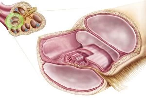
Background imageMagnification Collection: Medical illustration of endolymph in the membranous labyrinth of the inner ear

Medical illustration of endolymph in the membranous labyrinth of the inner ear
Medical ilustration showing endolymph in the membranous labyrinth of the inner ear

Background imageMagnification Collection: Microscopic view of histoplasmosis

Microscopic view of histoplasmosis. Histoplasmosis is an infection in the lungs caused by inhaling the spores of a fungus. This fungus, called Histoplasma capsulatum

Background imageMagnification Collection: Conceptual image of the Human Papilloma Virus

Conceptual image of the Human Papilloma Virus (HPV). HPV is a virus from the papillomavirus family that affects human skin and the moist membranes that line the body

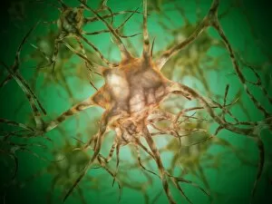
Background imageMagnification Collection: Microscopic view of multiple nerve cells, known as neurons

Microscopic view of multiple nerve cells, known as neurons
Microscopic view of multiple nerve cells, which are also called neurons. These are responsible for passing information around the central nervous system within the human body

Background imageMagnification Collection: Microscopic view of diplococcus bacterium

Microscopic view of diplococcus bacterium

Background imageMagnification Collection: Conceptual image of human eye and skull

Conceptual image of human eye and skull

Background imageMagnification Collection: Conceptual image of cell surface receptors

Conceptual image of cell surface receptors. Cell surface receptors are specialized integral membrane proteins that take part in communication between the cell and the outside world

Background imageMagnification Collection: Blood flow through a relaxed artery verus an artery in spasm

Blood flow through a relaxed artery verus an artery in spasm

Background imageMagnification Collection: Trachea cross-section showing normal and asthmatic bronchiole

Trachea cross-section showing normal and asthmatic bronchiole
Trachea cross-section showing comparison of normal and asthmatic bronchiole


All Professionally Made to Order for Quick Shipping

Magnification: Revealing the Hidden World From the intricate patterns of a snowflake to the delicate structure of a human cell, magnification allows us to explore and unravel the mysteries of our world. Peering through the lens, we uncover secrets that would otherwise remain unseen. Take for instance Crysotile asbestos, its fibrous nature exposed under intense magnification, reminding us of its hidden dangers lurking within. In medical illustrations, an appendix with appendicitis comes alive as we zoom in closer. The inflamed tissues become vividly apparent, highlighting the urgency for surgical intervention. Moving deeper into our bodies, we discover the liver - a complex organ responsible for countless vital functions. Under magnification, its intricate network of cells and blood vessels reveals itself like an elaborate cityscape bustling with activity. But it's not just within ourselves that magnification unveils wonders; it extends beyond to nature's realm. A dandelion's fruiting head transforms into a mesmerizing display when observed up close – each tiny seed ready to embark on its own journey through wind and time. Fractals captivate our imagination as well – from Julia fractals with their infinite complexity to Mandelbrot fractals' self-repeating patterns. These mathematical marvels remind us of nature's innate ability to create beauty even in abstract forms. Anton van Leeuwenhoek revolutionized science by observing animalcules through his microscope in 1795. His discoveries opened up new frontiers in microbiology and forever changed our understanding of life at microscopic levels. Even seemingly ordinary creatures like snails hold surprises when examined closely; their teeth reveal intricate structures designed for efficient feeding and survival. Ponds teem with life too - water fleas gracefully glide alongside green algae like Volvox aureus. Magnified images transport us into this miniature ecosystem where every organism plays a crucial role in maintaining balance and harmony. And finally, we come full circle with snowflakes.