Fraud Blocker Skip to main content

Mandible Collection (page 4)

The mandible, also known as the jawbone, is a fascinating and crucial component of our skull anatomy

Background imageMandible Collection: Anatomy of human skull, eyeball and arteries to brain

Anatomy of human skull, eyeball and arteries to brain

Background imageMandible Collection: A Collection of Coleoptera found in Chile, illustration from Historia de Chile

A Collection of Coleoptera found in Chile, illustration from Historia de Chile
XZL150467 A Collection of Coleoptera found in Chile, illustration from Historia de Chile, engraved by Lebrun, 1854 (litho) by French School, (19th century); Private Collection; French

Background imageMandible Collection: USA, Florida, STA 5 Water Management District, Birds, Black Skimmer, Flying and Skimming

USA, Florida, STA 5 Water Management District, Birds, Black Skimmer, Flying and Skimming and reflected in water

Background imageMandible Collection: Fragment of hippopotamus mandible and tusk; hyena mandible, from Liguria Region, Italy

Fragment of hippopotamus mandible and tusk; hyena mandible, from Liguria Region, Italy
Fossils - Fragment of hippopotamus mandible and tusk; hyena mandible. From Liguria Region

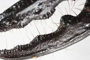
Background imageMandible Collection: Odontolabis cuvera (Stag Beetle) mandible, extreme close-up

Odontolabis cuvera (Stag Beetle) mandible, extreme close-up

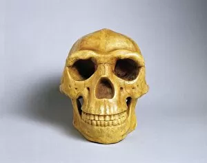
Background imageMandible Collection: Reconstruction of Peking Mans (Homo erectus pekinensis or Sinanthropus pekinensis)

Reconstruction of Peking Mans (Homo erectus pekinensis or Sinanthropus pekinensis) skull with mandible
Prehistory, China. Reconstruction of Peking Mans (Homo erectus pekinensis or Sinanthropus pekinensis) skull with mandible. Replica by Franz Weidenreich

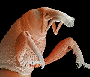
Background imageMandible Collection: Grain weevil

Grain weevil. Coloured scanning electron micrograph of the thorax and head of a grain weevil (Sitophilus granarius). This beetle infests and damages stored grain

Background imageMandible Collection: Red flour beetle

Red flour beetle. Coloured scanning electron micrograph (SEM) of a red flour beetle (Tribolium castaneum). One of the most significant pests of dried foods and the milling industry

Background imageMandible Collection: Grain weevil Z330 / 0408

Grain weevil Z330 / 0408
Grain weevil. Coloured scanning electron micrograph of a grain weevil (Sitophilus granarius). This beetle infests and damages stored grain, using the mandibles at the end of its elongated snout

Background imageMandible Collection: Human skull, artwork F007 / 7257

Human skull, artwork F007 / 7257
Human skull, computer artwork

Background imageMandible Collection: Human skull, artwork F007 / 7258

Human skull, artwork F007 / 7258
Human skull, computer artwork

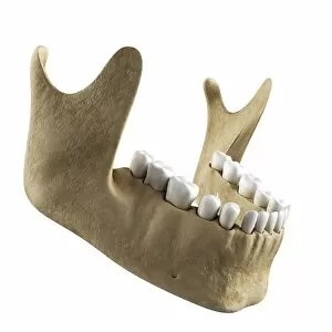
Background imageMandible Collection: Jawbone, artwork F007 / 7250

Jawbone, artwork F007 / 7250
Jawbone, computer artwork

Background imageMandible Collection: Human skull, artwork F007 / 7256

Human skull, artwork F007 / 7256
Human skull, computer artwork

Background imageMandible Collection: Lower jaw bone, artwork F006 / 3046

Lower jaw bone, artwork F006 / 3046
Lower jaw bone. Computer artwork showing the mandible bone

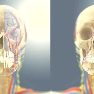
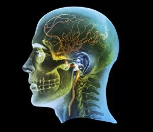
Background imageMandible Collection: Head and neck anatomy, artwork C014 / 0511

Head and neck anatomy, artwork C014 / 0511
Head and neck anatomy, computer artwork. Arteries are red, veins are blue, nerves are deep yellow and lymph vessels are bright yellow

Background imageMandible Collection: Neck muscle anatomy, 1831 artwork

Neck muscle anatomy, 1831 artwork
Neck muscle anatomy. Various views of the anatomy of the muscles of the neck. The main artwork shows the neck seen from the front

Background imageMandible Collection: Historical dentistry equipment

Historical dentistry equipment. 1940s model of a human head and jaw

Background imageMandible Collection: Caterpillar head

Caterpillar head. Close-up of the head of a small white caterpillar (order Lepidoptera), showing its mouthparts. Magnification: x28 when printed 10 centimetres wide

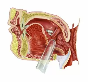
Background imageMandible Collection: Tonsils, artwork

Tonsils, artwork
Tonsils. Artwork of a section through the mouth showing a palatine tonsil. The clear rod points to the tonsillar fossa, a depression that is occupied by a palatine tonsil (round, right)

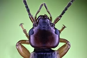
Background imageMandible Collection: Earwig head

Earwig head. Close-up of the head of an earwig (order Dermaptera), showing its compound eyes (centre left and right) and mouthparts (upper centre). Magnification: x17, when printed 10 centimetres wide

Background imageMandible Collection: Human head, MRI and 3D CT scans C016 / 6397

Human head, MRI and 3D CT scans C016 / 6397
Human head. Coloured composite image of a magnetic resonance imaging (MRI) scan of the brain and a 3D computed tomography (CT) scan of the skull of a 35 year old patient

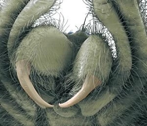
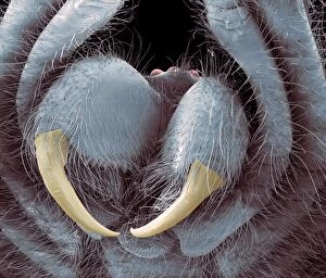
Background imageMandible Collection: Wolf spider jaws, SEM C016 / 9096

Wolf spider jaws, SEM C016 / 9096
Wolf spider jaws. Coloured scanning electron micrograph (SEM) of the head of a wolf spider (family Lycosidae), showing its large jaws (beige). Magnification: x16, when printed 10 centimetres wide

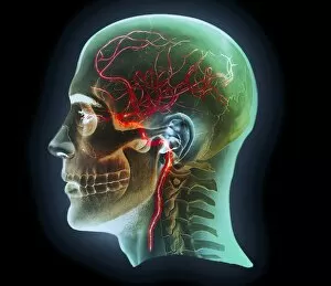
Background imageMandible Collection: Internal carotid artery, 3D CT scan C016 / 6399

Internal carotid artery, 3D CT scan C016 / 6399
Internal carotid artery. Coloured 3D computed tomography (CT) angiogram (blood vessel scan) of the head showing a normal internal carotid artery (red, in neck) and its intra-cerebral branches

Background imageMandible Collection: Internal carotid artery, 3D CT scan C016 / 6400

Internal carotid artery, 3D CT scan C016 / 6400
Internal carotid artery. Coloured 3D computed tomography (CT) angiogram (blood vessel scan) of the head showing a normal internal carotid artery (red, in neck) and its intra-cerebral branches

Background imageMandible Collection: Human head, MRI and 3D CT scans C016 / 6396

Human head, MRI and 3D CT scans C016 / 6396
Human head. Coloured composite image of a magnetic resonance imaging (MRI) scan of the brain and a 3D computed tomography (CT) scan of the skull of a 35 year old patient

Background imageMandible Collection: Human head, MRI and 3D CT scans C016 / 6398

Human head, MRI and 3D CT scans C016 / 6398
Human head. Coloured composite image of a magnetic resonance imaging (MRI) scan of the brain and a 3D computed tomography (CT) scan of the skull of a 35 year old patient

Background imageMandible Collection: Human head, MRI and 3D CT scans C016 / 6395

Human head, MRI and 3D CT scans C016 / 6395
Human head. Coloured composite image of a magnetic resonance imaging (MRI) scan of the brain and a 3D computed tomography (CT) scan of the skull of a 35 year old patient

Background imageMandible Collection: Wolf spider jaws, SEM C016 / 9095

Wolf spider jaws, SEM C016 / 9095
Wolf spider jaws. Coloured scanning electron micrograph (SEM) of the head of a wolf spider (family Lycosidae), showing its large jaws (beige). Magnification: x16, when printed 10 centimetres wide

Background imageMandible Collection: Human head, MRI and 3D CT scans C016 / 6494

Human head, MRI and 3D CT scans C016 / 6494
Human head. Coloured composite image of a magnetic resonance imaging (MRI) scan of the brain and a 3D computed tomography (CT) scan of the face of a 40 year old patient

Background imageMandible Collection: Tarantula spider, SEM

Tarantula spider, SEM
Tarantula spider. Coloured scanning electron micrograph (SEM) of the head of a Brazilian white striped bird-eating spider (Nhandu chromatus), showing its large mandibles (jaws, centre)
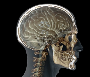
Background imageMandible Collection: Normal skull, 3D CT scan C016 / 6330

Normal skull, 3D CT scan C016 / 6330
Normal skull. Coloured cut-away 3D computed tomography (CT) scan of the healthy skull of a 35 year old patient

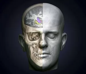
Background imageMandible Collection: Human head, 3D CT scan C016 / 6339

Human head, 3D CT scan C016 / 6339
Human head. Coloured 3D computed tomography (CT) scan of a healthy human head. The soft tissues are shown at right, the skull is seen at left

Background imageMandible Collection: Normal head and neck, 3D CT scan C016 / 6335

Normal head and neck, 3D CT scan C016 / 6335
Normal head. Coloured 3D computed tomography (CT) scan of the healthy head and neck of a 35 year old patient

Background imageMandible Collection: Normal head and neck, 3D CT scan C016 / 6334

Normal head and neck, 3D CT scan C016 / 6334
Normal head. Coloured 3D computed tomography (CT) scan of the healthy head and neck of a 35 year old patient

Background imageMandible Collection: Normal head and neck, MRI and 3D CT scans C016 / 6338

Normal head and neck, MRI and 3D CT scans C016 / 6338
Normal head. Coloured composite image of a magnetic resonance imaging (MRI) scan of the brain and a 3D computed tomography (CT) scan of the head and neck of a 35 year old patient

Background imageMandible Collection: Normal head and neck, MRI and 3D CT scans C016 / 6336

Normal head and neck, MRI and 3D CT scans C016 / 6336
Normal head. Coloured composite image of a magnetic resonance imaging (MRI) scan of the brain and a 3D computed tomography (CT) scan of the head and neck of a 35 year old patient

Background imageMandible Collection: Normal skull, 3D CT scan C016 / 6328

Normal skull, 3D CT scan C016 / 6328
Normal skull. Coloured 3D computed tomography (CT) scan of the healthy skull of a 35 year old patient

Background imageMandible Collection: Normal skull, 3D CT scan C016 / 6326

Normal skull, 3D CT scan C016 / 6326
Normal skull. Coloured 3D computed tomography (CT) scan of the healthy skull of a 35 year old patient

Background imageMandible Collection: Normal skull, 3D CT scan C016 / 6331

Normal skull, 3D CT scan C016 / 6331
Normal skull. Coloured cut-away 3D computed tomography (CT) scan of the healthy skull of a 35 year old patient

Background imageMandible Collection: Normal skull, 3D CT scan C016 / 6327

Normal skull, 3D CT scan C016 / 6327
Normal skull. Coloured 3D computed tomography (CT) scan of the healthy skull of a 35 year old patient

Background imageMandible Collection: Human head, 3D CT scan C016 / 6377

Human head, 3D CT scan C016 / 6377
Human head. Coloured 3D computed tomography (CT) scan of a profile view of a 47 year old patients head. The brain is purple and bone is light brown. At centre, behind the face, are the sinuses

Background imageMandible Collection: Stag beetle C016 / 2181

Stag beetle C016 / 2181
Stag beetle

Background imageMandible Collection: Stag beetle C016 / 2180

Stag beetle C016 / 2180
Stag beetle. View of the upper side of a male Lucanus cervus stag beetle. This insect lives in holes in old trees and dead trunks

Background imageMandible Collection: Stag beetle C016 / 2182

Stag beetle C016 / 2182
Stag beetle. Male Lucanus cervus stag beetle on a plant with seed pods. This insect lives in holes in old trees and dead trunks

Background imageMandible Collection: Stag beetle C016 / 2178

Stag beetle C016 / 2178
Stag beetle (Lucanus barbarossa). Specimen obtained from the University of Valencia, Spain

Background imageMandible Collection: Stag beetle C016 / 2213

Stag beetle C016 / 2213
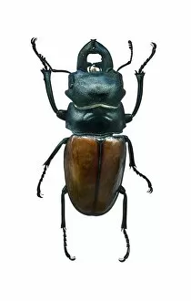
Stag beetle. View of the upper side of a male stag beetle (Odontolabis castelnaudi). Specimen obtained from the University of Valencia, Spain

Background imageMandible Collection: Benign dental tumour, CT scans C018 / 0589

Benign dental tumour, CT scans C018 / 0589
Benign dental tumour. Coloured computed tomography (CT) scans of the lower jaw of a 35 year old patient with a cemento-ossifying fibroma (purple)

Background imageMandible Collection: Jaw tumour, CT scan C018 / 0565

Jaw tumour, CT scan C018 / 0565
Jaw tumour. Coloured 3D computed tomography (CT) scan of the jaw of a 35 year old patient showing a benign (non-cancerous) bony lesion (indicated by arrows) in the mandible (lower jaw)


All Professionally Made to Order for Quick Shipping

The mandible, also known as the jawbone, is a fascinating and crucial component of our skull anatomy. Leonardo da Vinci's detailed sketches in his "Skull Anatomy" showcase the intricacies of this bone. A panoramic dental X-ray allows us to examine the mandible's structure and its relationship with teeth. Looking back in time, the Paranthropus boisei (Zinjanthropus) cranium (OH5) provides valuable insights into our evolutionary history. The study of human skulls through X-rays reveals not only the mandible but also other vital structures within our head. Nature never fails to amaze us; even tiny creatures like the red-barbed ant can be observed under a scanning electron microscope (SEM), revealing their unique mandibles adapted for various tasks. Meanwhile, a cross-section diagram illustrates how our mouth and jaw work together harmoniously. Insects are no exception when it comes to showcasing intriguing mandibular adaptations. Witnessing a red flour beetle in flight or examining an artwork depicting a Black Death rat flea reminds us of nature's diversity and complexity. However, sometimes unfortunate incidents occur that affect this essential bone. Fractured jawbones captured by X-rays remind us of both medical challenges and advancements in treating such injuries. Zooming further into nature's wonders, we encounter spiny spiders under SEMs - their menacing-looking yet functional jaws ready for capturing prey. Ultimately, whether studying ancient fossils or observing modern-day organisms, understanding the mandible plays an integral role in comprehending head and neck anatomy as depicted beautifully through artworks dedicated to this subject matter.