Fraud Blocker Skip to main content

Micro Organisms Collection (page 6)

Microorganisms, the tiny wonders of life that exist all around us, are a fascinating subject to explore

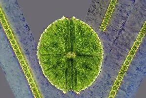
Background imageMicro Organisms Collection: Desmids, light micrograph

Desmids, light micrograph
Desmids. Phase contrast light micrograph of Micrasterias sp. and Hyalotheca sp. desmid green algae. Desmids are a common group of freshwater single-celled algae that have intricate cell walls

Background imageMicro Organisms Collection: Microalgae, light micrograph

Microalgae, light micrograph
Microalgae. Differential interference contrast (DIC) micrograph of Botryococcus braunii (round), and other species of green alga. Magnification: x278, when printed 10 centimetres wide

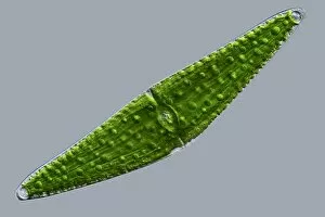
Background imageMicro Organisms Collection: Closterium desmid, light micrograph

Closterium desmid, light micrograph
Closterium desmid. Differential interference contrast (DIC) micrograph of a Closterium sp. desmid green alga. Desmids are a common group of freshwater single-celled algae that have intricate cell

Background imageMicro Organisms Collection: Haematococcus algae, light micrograph

Haematococcus algae, light micrograph
Haematococcus algae. Differential interference contrast (DIC) micrograph of a colony of Haematococcus sp. green alga. Magnification: x220, when printed 10 centimetres wide

Background imageMicro Organisms Collection: Ciliate protozoan, light micrograph

Ciliate protozoan, light micrograph
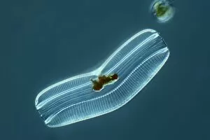
Ciliate protozoan. Darkfield illuminated light micrograph of a ciliate protozoan, showing green algae (green) that it has ingested

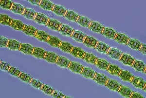
Background imageMicro Organisms Collection: Bambusina desmids, light micrograph

Bambusina desmids, light micrograph
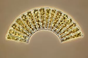
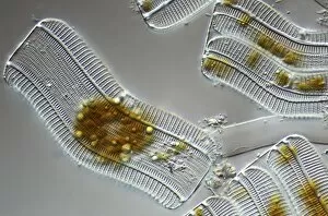
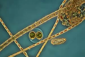
Bambusina desmids. Polarised light micrograph of a colony of Bambusina sp. desmid green algae. Desmids are a common group of freshwater single-celled algae that have intricate cell walls

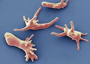
Background imageMicro Organisms Collection: Amoeba protozoa, SEM

Amoeba protozoa, SEM
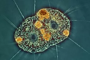
Amoeba protozoa. Coloured scanning electron micrograph (SEM) of four Amoeba sp. protozoa, showing their numerous pseudopodia (cytoplasmic extensions, leg-like)

Background imageMicro Organisms Collection: Rotifer, light micrograph

Rotifer, light micrograph
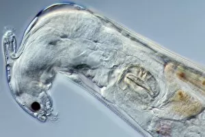
Rotifer. Darkfield illuminated light micrograph of a rotifer (phylum Rotifera). Rotifers (phylum Rotifera) are microscopic aquatic animals that are related to roundworms

Background imageMicro Organisms Collection: Prokaryote cell, artwork C016 / 6263

Prokaryote cell, artwork C016 / 6263
Prokaryote cell. Computer artwork showing the internal structure of a typical prokaryotic cell. Prokaryotes are organisms whose cells lack a membrane-bound nucleus (karyon)

Background imageMicro Organisms Collection: Prokaryote and eukaryote cells, artwork C016 / 6259

Prokaryote and eukaryote cells, artwork C016 / 6259
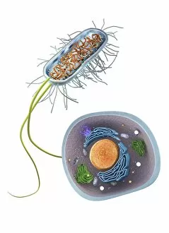
Prokaryote and eukaryote cells. Computer artwork comparing typical prokaryotic (top) and eukaryotic (bottom) cells. Prokaryotes are organisms whose cells lack a membrane-bound nucleus (karyon)

Background imageMicro Organisms Collection: Scenedesmus algae, SEM C014 / 1462

Scenedesmus algae, SEM C014 / 1462
Scenedesmus algae. Coloured scanning electron micrograph (SEM) of Scenedesmus sp. algae. Scenedesmus is a nonmotile colonial green algae that has its cells arranged in a row

Background imageMicro Organisms Collection: E. coli bacteria, SEM C014 / 0385

E. coli bacteria, SEM C014 / 0385
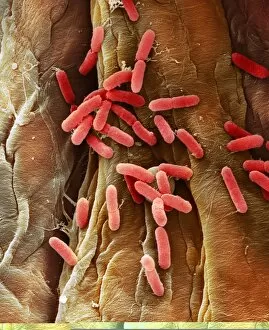
E. coli bacteria. Coloured scanning electron micrograph (SEM) of Escherichia coli (E. coli) bacteria (rod-shaped) found in a urine sample from a patient with a urinary tract infection (UTI)

Background imageMicro Organisms Collection: E. coli bacteria, SEM C014 / 0386

E. coli bacteria, SEM C014 / 0386
E. coli bacteria. Coloured scanning electron micrograph (SEM) of Escherichia coli (E. coli) bacteria (rod-shaped) found in a urine sample from a patient with a urinary tract infection (UTI)

Background imageMicro Organisms Collection: Diatoms, light micrograph C014 / 4675

Diatoms, light micrograph C014 / 4675
Diatoms. Differential interference contrast micrograph of a group of Meridion circulare diatoms. Diatoms are a group of photosynthetic, single-celled algae containing about 10, 000 species

Background imageMicro Organisms Collection: Diatom, light micrograph C014 / 4677

Diatom, light micrograph C014 / 4677
Diatom. Differential interference contrast micrograph of a Campylodiscus sp. diatom. Diatoms are a group of photosynthetic, single-celled algae containing about 10, 000 species

Background imageMicro Organisms Collection: Green hydra, light micrograph C014 / 4680

Green hydra, light micrograph C014 / 4680
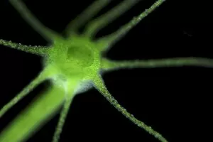
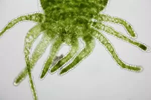
Green hydra. Darkfield illuminated light micrograph of the tentacled head of a green hydra (Hydra viridis). Hydra are small simple predatory fresh-water animals, belonging to the phylum Cnidaria

Background imageMicro Organisms Collection: Penicillium colony C014 / 4861

Penicillium colony C014 / 4861
Penicillium colony. Colony of Penicillium chrysogenum fungus growing on agar in a petri dish. This fungus produces the antibiotic penicillin G, seen as small droplets on the surface

Background imageMicro Organisms Collection: Green algae, light micrograph C014 / 4670

Green algae, light micrograph C014 / 4670
Green algae. Differential interference contrast micrograph of a single green alga, showing the chloroplast (green) inside. Algae use the chloroplasts to produce food through photosynthesis

Background imageMicro Organisms Collection: Diatoms, light micrograph C014 / 4671

Diatoms, light micrograph C014 / 4671
Diatoms. Differential interference contrast micrograph of a group of Achnantes longipes diatoms. Diatoms are a group of photosynthetic, single-celled algae containing about 10, 000 species

Background imageMicro Organisms Collection: Green hydra, light micrograph C014 / 4681

Green hydra, light micrograph C014 / 4681
Green hydra. Darkfield illuminated light micrograph of a green hydra (Hydra viridis), showing its tentacled head. Hydra are small simple predatory fresh-water animals

Background imageMicro Organisms Collection: Anabaena algae, SEM

Anabaena algae, SEM
Anabaena algae. Coloured scanning electron micrograph (SEM) of Anabaena sp. freshwater alga from a pond. Anabaena is a filamentous species of blue-green algae (cyanobacteria)

Background imageMicro Organisms Collection: Diatom, light micrograph C014 / 4669

Diatom, light micrograph C014 / 4669
Diatom. Differential interference contrast micrograph of an Achnantes longipes diatom. Diatoms are a group of photosynthetic, single-celled algae containing about 10, 000 species

Background imageMicro Organisms Collection: Radiolaria, light micrograph C014 / 4672

Radiolaria, light micrograph C014 / 4672
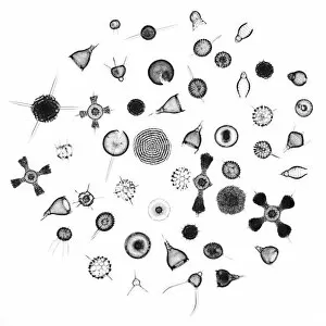
Radiolaria. Collection of light micrographs showing various different species of radiolaria. Radiolaria (or radiolarians) are amoeboid protozoa that produce intricate mineral skeletons

Background imageMicro Organisms Collection: Penicillium colony C014 / 4857

Penicillium colony C014 / 4857
Penicillium colony. Colony of Penicillium chrysogenum fungus growing on agar in a petri dish. This fungus produces the antibiotic penicillin G, seen as small droplets on the surface

Background imageMicro Organisms Collection: Diatoms, light micrograph C014 / 4668

Diatoms, light micrograph C014 / 4668
Diatoms. Differential interference contrast micrograph of a group of Achnantes longipes diatoms. Diatoms are a group of photosynthetic, single-celled algae containing about 10, 000 species

Background imageMicro Organisms Collection: Diatoms, light micrograph C014 / 4674

Diatoms, light micrograph C014 / 4674
Diatoms. Differential interference contrast micrograph of a group of Achnantes longipes diatoms. Diatoms are a group of photosynthetic, single-celled algae containing about 10, 000 species

Background imageMicro Organisms Collection: Green hydra, light micrograph C014 / 4678

Green hydra, light micrograph C014 / 4678
Green hydra. Light micrograph of the tentacled head of a green hydra (Hydra viridis). Hydra are small simple predatory fresh-water animals, belonging to the phylum Cnidaria

Background imageMicro Organisms Collection: Ciliate protozoan, light micrograph C014 / 4676

Ciliate protozoan, light micrograph C014 / 4676
Ciliate protozoan. Differential interference contrast micrograph of a ciliate protozoan, showing the symbiotic green algae (Zoochlorellae, green) contained in vacuoles within

Background imageMicro Organisms Collection: Rotifer, light micrograph C014 / 4666

Rotifer, light micrograph C014 / 4666
Rotifer, differential interference contrast micrograph. These animals are related to roundworms. They are usually freshwater organisms though a few marine sp

Background imageMicro Organisms Collection: Green hydra, light micrograph C014 / 4679

Green hydra, light micrograph C014 / 4679
Green hydra. Darkfield illuminated light micrograph of a green hydra (Hydra viridis), showing its tentacled head. Hydra are small simple predatory fresh-water animals

Background imageMicro Organisms Collection: Pediastrum green algae, micrograph

Pediastrum green algae, micrograph
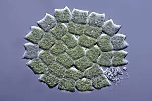
Pediastrum green algae. Differential interference contrast light micrograph of a circular colony of Pediastrum sp. green algae (Chlorophyte). Pediastrum is a type of freshwater colonial algae

Background imageMicro Organisms Collection: Ciliate protozoan, light micrograph C014 / 4667

Ciliate protozoan, light micrograph C014 / 4667
Ciliate protozoan. Differential interference contrast micrograph of a ciliate protozoan, showing the symbiotic green algae (Zoochlorellae, green) contained in vacuoles within

Background imageMicro Organisms Collection: Hand bacteria C014 / 4839

Hand bacteria C014 / 4839
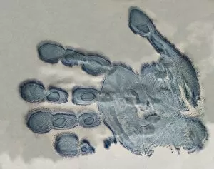
Hand bacteria. Colony of bacteria that have grown where a hand was pressed onto an agar plate

Background imageMicro Organisms Collection: Bdelloid rotifer, SEM

Bdelloid rotifer, SEM
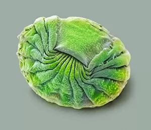
Bdelloid rotifer. Coloured scanning electron micrograph (SEM) of a rotifer (class Bdelloidea) from a pond. Rotifers (phylum Rotifera) are microscopic aquatic animals that are related to roundworms

Background imageMicro Organisms Collection: Foraminiferan

Foraminiferan, darkfield light micrograph. Foraminifera are marine single-celled protozoa that construct and inhabit shells composed of several chambers

Background imageMicro Organisms Collection: E. coli DNA mismatch repair complex

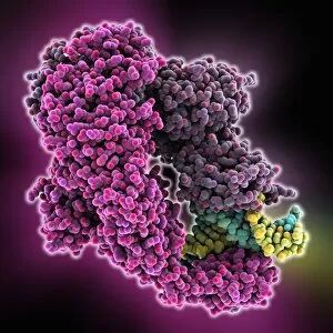
E. coli DNA mismatch repair complex. Computer model showing the structure of a MutS (Mutator S) protein from Escherichia coli (E)

Background imageMicro Organisms Collection: Closterium desmid, light micrograph C016 / 8608

Closterium desmid, light micrograph C016 / 8608
Closterium desmid. Interphase contrast micrograph of a Closterium sp. desmid green alga. Desmids are a common group of freshwater single-celled algae that have intricate cell walls

Background imageMicro Organisms Collection: Amoeba protozoa, light micrograph C016 / 8607

Amoeba protozoa, light micrograph C016 / 8607
Amoeba protozoa. Interphase contrast light micrograph of two Amoeba sp. protozoa eating each other, showing its numerous pseudopodia (cytoplasmic extensions, leg-like)

Background imageMicro Organisms Collection: Diatom, light micrograph C016 / 8606

Diatom, light micrograph C016 / 8606
Diatom. Phase-contrast light micrograph of a diatom from the Mediterranean. Diatoms are a group of photosynthetic, single-celled algae containing about 10, 000 species

Background imageMicro Organisms Collection: Diatoms, light micrograph C016 / 8605

Diatoms, light micrograph C016 / 8605
Diatoms, Shearing contrast light micrograph. Diatoms are a group of photosynthetic, single-celled algae containing about 10, 000 species

Background imageMicro Organisms Collection: Diatoms, light micrograph C016 / 8598

Diatoms, light micrograph C016 / 8598
Diatoms. Interphase contrast light micrograph of a selection of differently shaped diatoms. Diatoms are a group of photosynthetic, single-celled algae containing about 10, 000 species

Background imageMicro Organisms Collection: Diatoms, light micrograph C016 / 8602

Diatoms, light micrograph C016 / 8602
Diatoms. Phase-contrast light micrograph of diatoms from the Mediterranean. Diatoms are a group of photosynthetic, single-celled algae containing about 10, 000 species

Background imageMicro Organisms Collection: Foraminifera, light micrograph C016 / 8597

Foraminifera, light micrograph C016 / 8597
Foraminifera. Interphase contrast light micrograph of a selection of different foraminifera. Foraminifera are marine single-celled protozoa that construct

Background imageMicro Organisms Collection: Diatoms, light micrograph C016 / 8600

Diatoms, light micrograph C016 / 8600
Diatoms. Differential interference contrast (DIC) light micrograph of diatoms from the Mediterranean. Diatoms are a group of photosynthetic, single-celled algae containing about 10, 000 species

Background imageMicro Organisms Collection: Green algae, light micrograph C016 / 8595

Green algae, light micrograph C016 / 8595
Green algae, Interphase contrast light micrograph. Magnification: x280, when printed 10 centimetres wide

Background imageMicro Organisms Collection: Ciliate protozoan, light micrograph C016 / 8593

Ciliate protozoan, light micrograph C016 / 8593
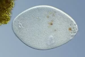
Ciliate protozoan. Differential interference contrast (DIC) light micrograph of a ciliate protozoan. Magnification: x280 when printed 10 centimetres wide

Background imageMicro Organisms Collection: Hypotrich protozoan, light micrograph C016 / 8592

Hypotrich protozoan, light micrograph C016 / 8592
Hypotrich protozoan. Differential interference contrast (DIC) light micrograph of a Euplotes sp. hypotrich ciliate protozoan. Magnification: x280 when printed 10 centimetres wide

Background imageMicro Organisms Collection: Fossil diatom, light micrograph C016 / 8586

Fossil diatom, light micrograph C016 / 8586
Fossil diatom. Interphase contrast light micrograph of a fossilised diatom. Diatoms are a group of photosynthetic, single-celled algae containing about 10, 000 species


All Professionally Made to Order for Quick Shipping

Microorganisms, the tiny wonders of life that exist all around us, are a fascinating subject to explore. Under the lens of a scanning electron microscope (SEM), we can witness their intricate structures and unravel their hidden secrets. Take E. Coli bacteria for example; when magnified through an SEM, they reveal their rod-shaped bodies with flagella protruding from one end. These microscopic creatures play crucial roles in our digestive system but can also cause infections if not properly handled. Similarly, Salmonella bacteria appear as elongated cells under SEM, reminding us of the importance of proper food handling and hygiene practices to prevent contamination. The colored transmission electron microscopy (TEM) image of Yersinia pestis bacteria showcases its unique features that were responsible for devastating outbreaks like the infamous Black Death. Switching gears to fungi, Candida fungus is captured beautifully in an SEM image displaying its filamentous structure. This opportunistic pathogen can cause infections in immunocompromised individuals and highlights the need for effective antifungal treatments. Delving into history, we encounter anthrax cultures depicted in a historical diagram. This bacterium has been weaponized throughout time due to its ability to form spores resistant to harsh conditions – a chilling reminder of humanity's dark side. Norovirus particles come into focus through TEM imagery; these small viral entities are notorious for causing gastroenteritis outbreaks worldwide and serve as a constant reminder about practicing good personal hygiene habits. Tuberculosis bacteria capture attention with their distinctive shape under SEM: slender rods often forming chains resembling delicate spirals. This ancient disease continues to pose significant health challenges globally despite medical advancements made over centuries. Streptomyces bacteria showcase their beauty by forming spiral spore chains visible even without high-powered microscopes. These remarkable organisms produce antibiotics vital for human health while maintaining ecological balance within soil ecosystems. Flagellate bacteria remind us that movement is not limited solely to larger organisms; these tiny creatures possess whip-like appendages that propel them through their microscopic habitats.