Fraud Blocker Skip to main content

Micro Organisms Collection (page 9)

Microorganisms, the tiny wonders of life that exist all around us, are a fascinating subject to explore

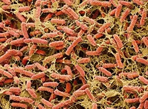
Background imageMicro Organisms Collection: Pond bacteria C015 / 6371

Pond bacteria C015 / 6371
Pond bacteria. Coloured scanning electron micrograph (SEM) of rod-shaped bacteria (bacilli) from a freshwater pond. Magnification: x4000, when printed 10cm wide

Background imageMicro Organisms Collection: Pond bacteria C015 / 6369

Pond bacteria C015 / 6369
Pond bacteria. Coloured scanning electron micrograph (SEM) of rod-shaped bacteria (bacilli) from a freshwater pond. Magnification: x4000, when printed 10cm wide

Background imageMicro Organisms Collection: Fossilised foraminiferan, SEM C015 / 5772

Fossilised foraminiferan, SEM C015 / 5772
Fossilised foraminiferan. Scanning electron micrograph (SEM) of a section through the fossilised shell of a foraminiferan in a sample of limestone

Background imageMicro Organisms Collection: Netrium desmids, light micrograph

Netrium desmids, light micrograph
Netrium desmids. Polarised light micrograph of Netrium sp. desmid green alga. Desmids are a common group of freshwater single-celled algae that have intricate cell walls

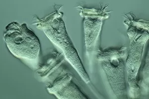
Background imageMicro Organisms Collection: Vorticella protozoan, light micrograph

Vorticella protozoan, light micrograph
Vorticella protozoan. Rheinberg illuminated light micrograph of a Vorticella sp. ciliate protozoan. This single-celled organism consists of a bell-shaped head

Background imageMicro Organisms Collection: Cosmarium desmids, light micrograph

Cosmarium desmids, light micrograph
Phase contrast, magnification: 200 X on APS-C sensor

Background imageMicro Organisms Collection: Golden algae, light micrograph

Golden algae, light micrograph
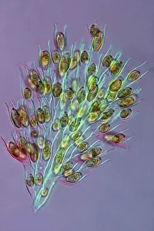
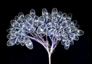
Golden algae. Polarised light micrograph of a colony of Dinobryon sp. golden algae (chrysophytes). Magnification: x200 when printed 10 centimetres wide

Background imageMicro Organisms Collection: Paramecium bursaria protozoan, light micr

Paramecium bursaria protozoan, light micr
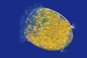
Paramecium bursaria protozoan, differential interference contrast light micrograph. This ciliate protozoan inhabits freshwater, where it feeds mainly on bacteria

Background imageMicro Organisms Collection: Euastrum desmids, light micrograph

Euastrum desmids, light micrograph
Euastrum desmids. Polarised light micrograph of Euastrum sp. desmid green algae. Desmids are a common group of freshwater single-celled algae that have intricate cell walls

Background imageMicro Organisms Collection: Blepharisma protozoan, light micrograph

Blepharisma protozoan, light micrograph
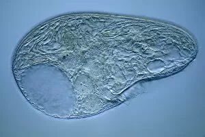
Blepharisma protozoan. Differential interference contrast micrograph of a Blepharisma japonicum protozoan. These ciliate protozoa move by beating their cilia

Background imageMicro Organisms Collection: Blepharisma protozoa, light micrograph

Blepharisma protozoa, light micrograph
Blepharisma protozoa. Differential interference contrast micrograph of Blepharisma japonicum protozoa. These ciliate protozoa move by beating their cilia

Background imageMicro Organisms Collection: Micrasterias desmids, light micrograph

Micrasterias desmids, light micrograph
Micrasterias desmids. Polarised light micrograph of Micrasterias truncata desmid green algae. Desmids are a common group of freshwater single-celled algae that have intricate cell walls

Background imageMicro Organisms Collection: MERS coronavirus, artwork C016 / 3611

MERS coronavirus, artwork C016 / 3611
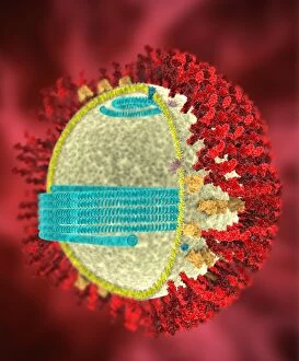
MERS coronavirus proteins. Cutaway computer model showing the protein structure of a MERS coronavirus particle (virion). MERS (Middle East respiratory syndrome)

Background imageMicro Organisms Collection: Plankton and fish, artwork C016 / 3448

Plankton and fish, artwork C016 / 3448
Plankton and fish. Artwork of plankton as viewed under a magnifying glass, with seabream (Diplodus vulgaris) fish

Background imageMicro Organisms Collection: E. coli induced cell death, SEM C016 / 3078

E. coli induced cell death, SEM C016 / 3078
E. coli induced cell death. Coloured scanning electron micrograph (SEM) of a macrophage white blood cell (centre) that is being destroyed by toxins released by Escherichia coli (E)

Background imageMicro Organisms Collection: E. coli induced cell death, SEM C016 / 3077

E. coli induced cell death, SEM C016 / 3077
E. coli induced cell death. Coloured scanning electron micrograph (SEM) of a macrophage white blood cell (centre) that is being destroyed by toxins released by Escherichia coli (E)

Background imageMicro Organisms Collection: SARS coronavirus proteins, artwork C016 / 3056

SARS coronavirus proteins, artwork C016 / 3056
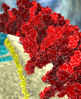
SARS coronavirus proteins. Computer model showing the spike proteins (red) of a SARS coronavirus particle (virion). SARS (severe acute respiratory syndrome)

Background imageMicro Organisms Collection: SARS coronavirus, artwork C016 / 3055

SARS coronavirus, artwork C016 / 3055
SARS coronavirus proteins. Computer artwork of SARS coronavirus particles (virions). SARS (severe acute respiratory syndrome)

Background imageMicro Organisms Collection: SARS coronavirus, artwork C016 / 3054

SARS coronavirus, artwork C016 / 3054
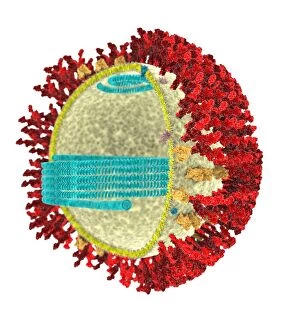
SARS coronavirus proteins. Cutaway computer model showing the protein structure of a SARS coronavirus particle (virion). SARS (severe acute respiratory syndrome)

Background imageMicro Organisms Collection: SARS coronavirus, artwork C016 / 3053

SARS coronavirus, artwork C016 / 3053
SARS coronavirus proteins. Cutaway computer model showing the protein structure of a SARS coronavirus particle (virion). SARS (severe acute respiratory syndrome)

Background imageMicro Organisms Collection: SARS coronavirus proteins, artwork C016 / 3052

SARS coronavirus proteins, artwork C016 / 3052
SARS coronavirus proteins. Computer model showing the proteins of a SARS coronavirus particle (virion). SARS (severe acute respiratory syndrome)

Background imageMicro Organisms Collection: Vorticella protozoa, light micrograph C016 / 3042

Vorticella protozoa, light micrograph C016 / 3042
Vorticella protozoa. Differential interference contrast (DIC) light micrograph of Vorticella sp. ciliate protozoa. These single-celled organisms consist of a bell-shaped head

Background imageMicro Organisms Collection: Vorticella protozoan, light micrograph C016 / 3043

Vorticella protozoan, light micrograph C016 / 3043
Vorticella protozoan. Differential interference contrast (DIC) light micrograph of a Vorticella sp. ciliate protozoan. This single-celled organism consists of a bell-shaped head

Background imageMicro Organisms Collection: Vorticella protozoa, light micrograph C016 / 3041

Vorticella protozoa, light micrograph C016 / 3041
Vorticella protozoa. Differential interference contrast (DIC) light micrograph of Vorticella sp. ciliate protozoa. These single-celled organisms consist of a bell-shaped head

Background imageMicro Organisms Collection: Vorticella protozoa, light micrograph C016 / 3040

Vorticella protozoa, light micrograph C016 / 3040
Vorticella protozoa. Differential interference contrast (DIC) light micrograph of Vorticella sp. ciliate protozoa. These single-celled organisms consist of a bell-shaped head

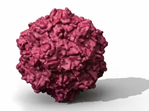
Background imageMicro Organisms Collection: Satellite tobacco necrosis virus C014 / 0005

Satellite tobacco necrosis virus C014 / 0005
Satellite tobacco necrosis virus. Computer artwork showing the surface structure of a tobacco necrosis satellivirus particle (virion)

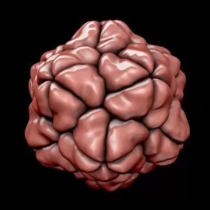
Background imageMicro Organisms Collection: Canine parvovirus particle C013 / 9966

Canine parvovirus particle C013 / 9966
Canine parvovirus particle. Computer artwork showing the structure of the outer protein coat (capsid) of a canine parvovirus type 2 particle (virion)

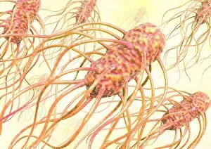
Background imageMicro Organisms Collection: Salmonella bacteria, artwork C013 / 7809

Salmonella bacteria, artwork C013 / 7809
Salmonella bacteria, computer artwork. Salmonella sp. bacteria are gram-negative rod-shaped bacteria that have flagella (hair-like structures) that they use for locomotion

Background imageMicro Organisms Collection: Salmonella bacteria, artwork C013 / 7808

Salmonella bacteria, artwork C013 / 7808
Salmonella bacteria, computer artwork. Salmonella sp. bacteria are gram-negative rod-shaped bacteria that have flagella (hair-like structures) that they use for locomotion

Background imageMicro Organisms Collection: Bird flu virus particle, artwork C013 / 4650

Bird flu virus particle, artwork C013 / 4650
Bird flu virus particle. Computer artwork showing the internal and external structures of an influenza (flu) A subtype H5N1 (bird flu) virus particle (virion)

Background imageMicro Organisms Collection: Ross River virus particle, artwork C013 / 4641

Ross River virus particle, artwork C013 / 4641
Ross River virus particle. Computer artwork showing the outer structure of a Ross River virus (RRV) particle (virion). RRV is a small encapsulated single-strand RNA alphavirus endemic to Australia

Background imageMicro Organisms Collection: Marburg virus, artwork C013 / 4633

Marburg virus, artwork C013 / 4633
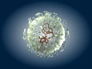
Marburg virus. Computer artwork of Marburg virus particles (virions). This tubular RNA (ribonucleic acid, coloured spheres) virus causes Marburg haemorrhagic fever in humans and non-human primates

Background imageMicro Organisms Collection: Hepatitis C virus, artwork

Hepatitis C virus, artwork
Hepatitis C virus. Computer artwork of hepatitis C virus particles (virions) in the human body. The virus consists of a core of RNA (ribonucleic acid) enclosed in a capsid

Background imageMicro Organisms Collection: EHEC E. coli bacteria, artwork C013 / 4621

EHEC E. coli bacteria, artwork C013 / 4621
EHEC E. coli bacteria. Computer artwork of a enterohaemorrhagic Escherichia coli (EHEC) bacteria in the human gut. E. coli bacteria are a normal part of the intestinal flora in humans

Background imageMicro Organisms Collection: Mould fungus, SEM

Mould fungus, SEM
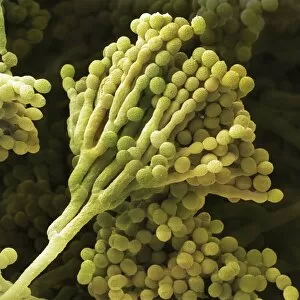
Mould fungus (Penicillium expansum), coloured scanning electron micrograph (SEM). This fungus, also called blue mould, is a pathogen of plant material

Background imageMicro Organisms Collection: Staphylococcus aureus bacteria, SEM C013 / 4771

Staphylococcus aureus bacteria, SEM C013 / 4771
Staphylococcus aureus bacteria. Coloured scanning electron micrograph (SEM) of Staphylococcus aureus bacteria (yellow). S

Background imageMicro Organisms Collection: Staphylococcus aureus bacteria, SEM C013 / 4770

Staphylococcus aureus bacteria, SEM C013 / 4770
Staphylococcus aureus bacteria. Coloured scanning electron micrograph (SEM) of Staphylococcus aureus bacteria (yellow). S

Background imageMicro Organisms Collection: Staphylococcus aureus bacteria, SEM C013 / 4769

Staphylococcus aureus bacteria, SEM C013 / 4769
Staphylococcus aureus bacteria. Coloured scanning electron micrograph (SEM) of Staphylococcus aureus bacteria (yellow). S

Background imageMicro Organisms Collection: Salmonella bacterium, artwork C013 / 4728

Salmonella bacterium, artwork C013 / 4728
Salmonella bacterium, computer artwork. Salmonella sp. bacteria are gram-negative rod-shaped bacteria that have flagella (hair-like structures) that they use for locomotion

Background imageMicro Organisms Collection: Salmonella bacteria, artwork C013 / 4727

Salmonella bacteria, artwork C013 / 4727
Salmonella bacteria, computer artwork. Salmonella sp. bacteria are gram-negative rod-shaped bacteria that have flagella (hair-like structures) that they use for locomotion

Background imageMicro Organisms Collection: Viral recognition by antibodies, artwork C013 / 4722

Viral recognition by antibodies, artwork C013 / 4722
Viral recognition by antibodies. Computer artwork of rabies (family Rhabdoviridae) virus particles (virions, pink) being identified by monoclonal antibodies (Y-shaped, blue)

Background imageMicro Organisms Collection: Rabies virus, artwork C013 / 4704

Rabies virus, artwork C013 / 4704
Rabies virus. Computer artwork of rabies (family Rhabdoviridae) virus particles (virions). The virus has a protein coat or capsid, which is made up of helically arranged subunits called capsomeres

Background imageMicro Organisms Collection: Rabies virus, artwork C013 / 4703

Rabies virus, artwork C013 / 4703
Rabies virus. Computer artwork of a rabies (family Rhabdoviridae) virus particle (virion). The virus has a protein coat or capsid, which is made up of helically arranged subunits called capsomeres

Background imageMicro Organisms Collection: Cabbage root infection, SEM

Cabbage root infection, SEM
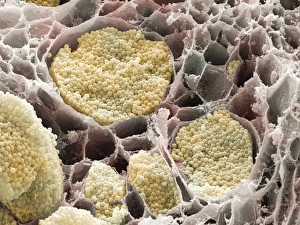
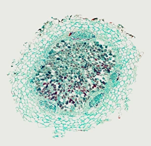
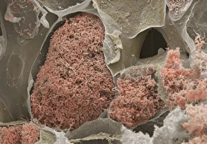
Cabbage root infection. Coloured scanning electron micrograph (SEM) of a cross-section through one of the roots of a cabbage (Brassica sp)

Background imageMicro Organisms Collection: LM of a root nodule

LM of a root nodule
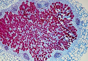
Light micrograph of a root nodule of the broad bean, Vicia faba, a leguminous plant. The nodule develops in response to invasion by the bacteria Rhizobium sp

Background imageMicro Organisms Collection: Nitrogen-fixing root nodule, micrograph

Nitrogen-fixing root nodule, micrograph
Nitrogen-fixing root nodule. Light micrograph of a section through a root nodule of a broad bean plant (Faba vulgaris) showing the symbiotic Rhizobium leguminosarum bacteria (purple) within it

Background imageMicro Organisms Collection: Nitrogen-fixing bacteria

Nitrogen-fixing bacteria. Coloured scanning electron micrograph (SEM) of Rhizobium leguminosarum nitrogen fixing bacteria (brown)


All Professionally Made to Order for Quick Shipping

Microorganisms, the tiny wonders of life that exist all around us, are a fascinating subject to explore. Under the lens of a scanning electron microscope (SEM), we can witness their intricate structures and unravel their hidden secrets. Take E. Coli bacteria for example; when magnified through an SEM, they reveal their rod-shaped bodies with flagella protruding from one end. These microscopic creatures play crucial roles in our digestive system but can also cause infections if not properly handled. Similarly, Salmonella bacteria appear as elongated cells under SEM, reminding us of the importance of proper food handling and hygiene practices to prevent contamination. The colored transmission electron microscopy (TEM) image of Yersinia pestis bacteria showcases its unique features that were responsible for devastating outbreaks like the infamous Black Death. Switching gears to fungi, Candida fungus is captured beautifully in an SEM image displaying its filamentous structure. This opportunistic pathogen can cause infections in immunocompromised individuals and highlights the need for effective antifungal treatments. Delving into history, we encounter anthrax cultures depicted in a historical diagram. This bacterium has been weaponized throughout time due to its ability to form spores resistant to harsh conditions – a chilling reminder of humanity's dark side. Norovirus particles come into focus through TEM imagery; these small viral entities are notorious for causing gastroenteritis outbreaks worldwide and serve as a constant reminder about practicing good personal hygiene habits. Tuberculosis bacteria capture attention with their distinctive shape under SEM: slender rods often forming chains resembling delicate spirals. This ancient disease continues to pose significant health challenges globally despite medical advancements made over centuries. Streptomyces bacteria showcase their beauty by forming spiral spore chains visible even without high-powered microscopes. These remarkable organisms produce antibiotics vital for human health while maintaining ecological balance within soil ecosystems. Flagellate bacteria remind us that movement is not limited solely to larger organisms; these tiny creatures possess whip-like appendages that propel them through their microscopic habitats.