Fraud Blocker Skip to main content

Microscope Collection (page 11)

"Unveiling the Unseen: Exploring the Microscopic World" Step into a realm of hidden wonders

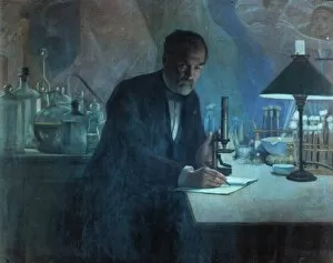
Background imageMicroscope Collection: LOUIS PASTEUR (1822-1895). French chemist and microbiologist. Pasteur in his laboratory

LOUIS PASTEUR (1822-1895). French chemist and microbiologist. Pasteur in his laboratory. Fresco by Fournier

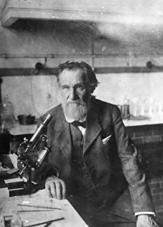
Background imageMicroscope Collection: ELIE METCHNIKOFF (1845-1916). Russian zoologist and bacteriologist

ELIE METCHNIKOFF (1845-1916). Russian zoologist and bacteriologist

Background imageMicroscope Collection: Male doctor using microscope in surgery, (B&W)

Male doctor using microscope in surgery, (B&W)

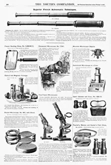
Background imageMicroscope Collection: SCIENTIFIC INSTRUMENTS, 1890. American magazine advertisements for various scientific instruments

SCIENTIFIC INSTRUMENTS, 1890. American magazine advertisements for various scientific instruments, 1890

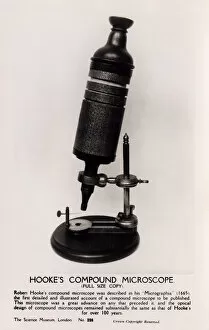
Background imageMicroscope Collection: Robert Hookes Microscope

Robert Hookes Microscope
Full-size copy of Robert Hookes Compound Microscope - held at the Science Museum, London. Hooke, an English natural philosopher, architect and polymath (1635-1703) - author of Micrographia (1665)

Background imageMicroscope Collection: Picture No. 11014617

Picture No. 11014617
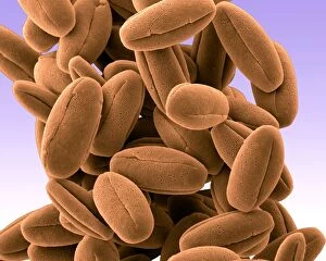
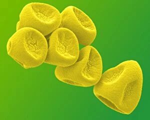
Veronica Pollen Scanning Electron Micrograph (SEM): Magnification x8220 (if print A4 size: 29.7 cm wide) - These small pollen grains are sticky and are carried away by pollinating insects Date:

Background imageMicroscope Collection: Picture No. 11014615

Picture No. 11014615
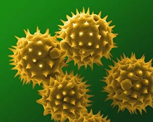
Sunflower Pollen Scanning Electron Micrograph (SEM): Magnification x10, 000 (if print A4 size: 29.7 cm wide) - The large flowers are insect pollinated

Background imageMicroscope Collection: Picture No. 11014607

Picture No. 11014607
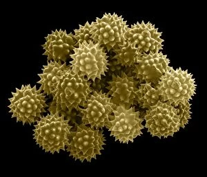
Groundsel Pollen Scanning Electron Micrograph (SEM): Magnification 2 x6940 (if print A4 size: 29.7 cm wide) - Groundsel is a common weed - It is insect pollinated; provides food for many insects

Background imageMicroscope Collection: Picture No. 11014606

Picture No. 11014606
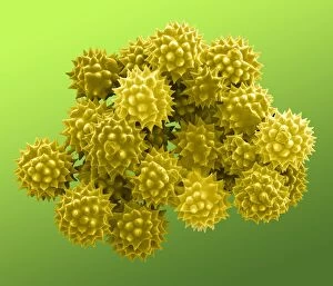
Groundsel Pollen Scanning Electron Micrograph (SEM): Magnification 2 x6940 (if print A4 size: 29.7 cm wide) - Groundsel is a common weed - It is insect pollinated; provides food for many insects

Background imageMicroscope Collection: Picture No. 11014604

Picture No. 11014604
Gorse Pollen Scanning Electron Micrograph (SEM): Magnification 1 x10, 000 (if print A4 size: 29.7 cm wide) - Gorse is a bush, with brigh yellow flowers in spring and early summer

Background imageMicroscope Collection: Picture No. 11014601

Picture No. 11014601
Daffodil Pollen Scanning Electron Micrograph (SEM): Magnification x8930 (if print A4 size: 29.7 cm wide) - Daffodils are popular and common springtime flowers. Insect pollinated Date:

Background imageMicroscope Collection: Picture No. 11014593

Picture No. 11014593
Peruvian Lily / Alstroemeria Pollen on Anther Scanning Electron Micrograph (SEM): Magnification x6520 (if print A4 size: 29.7 cm wide)

Background imageMicroscope Collection: Capturing the German dye trade (London Dye Manufacturing Co). A microscopic exmination

Capturing the German dye trade (London Dye Manufacturing Co). A microscopic exmination of colur crystals. 13 November 1920

Background imageMicroscope Collection: Picture No. 10873636

Picture No. 10873636
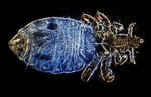
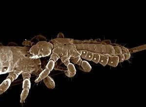
Human Head Louse (Pediculus humanus capitis) Date:

Background imageMicroscope Collection: Picture No. 10874130

Picture No. 10874130
Light Micrograph (LM): Hookworm (Ancylostoma duodenale) Date:

Background imageMicroscope Collection: Picture No. 10873581

Picture No. 10873581
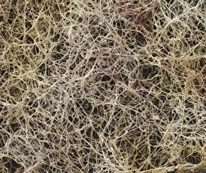
Athleteճ Foot Fungus (Tinea pedis) Date:

Background imageMicroscope Collection: Picture No. 10873643

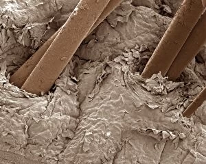
Picture No. 10873643
Human scalp Date:

Background imageMicroscope Collection: Picture No. 10873686

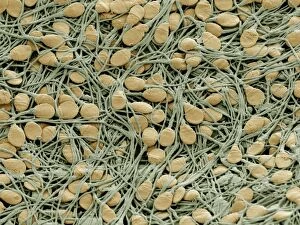
Picture No. 10873686
Human sperm cells Date:

Background imageMicroscope Collection: Picture No. 10873644

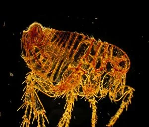
Picture No. 10873644
Human Flea - male (Pulex irritans) Date:

Background imageMicroscope Collection: Picture No. 10873600

Picture No. 10873600
Human Crab Louse (Phthirus pubis) Date:

Background imageMicroscope Collection: Picture No. 10873580

Picture No. 10873580
Athleteճ Foot Fungu (Tinea pedis) Date:

Background imageMicroscope Collection: Picture No. 10873601

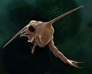
Picture No. 10873601
Crab larvae (Zoea larvae) Date:

Background imageMicroscope Collection: Picture No. 10873638

Picture No. 10873638
Human Head Louse (Pediculus humanus capitis) Date:

Background imageMicroscope Collection: Picture No. 10873634

Picture No. 10873634
Cochlea of Guinea Pig Date:

Background imageMicroscope Collection: Picture No. 10873615

Picture No. 10873615
Human Ovum in Fallopian Tube : This freshly fertilized egg passes down the Fallopian tube on its way to implantation in the wall of the uterus ( ) Date:

Background imageMicroscope Collection: Picture No. 10873617

Picture No. 10873617
Human finger tip print Date:

Background imageMicroscope Collection: Picture No. 10873675

Picture No. 10873675
Human White and Red Blood Cells Date:

Background imageMicroscope Collection: Picture No. 10873616

Picture No. 10873616
Human Ovum in Fallopian Tube : This freshly fertilized egg passes down the Fallopian tube on its way to implantation in the wall of the uterus Date:

Background imageMicroscope Collection: Picture No. 10873703

Picture No. 10873703
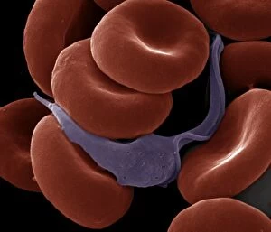
Sleeping Sickness Parasite in red blood cells (Trypanosoma sp.) Date:

Background imageMicroscope Collection: Picture No. 10873596

Picture No. 10873596
Human Cochlea Date:

Background imageMicroscope Collection: Picture No. 10891096

Picture No. 10891096
Paleontologist / Palaeontologist Eric Depre using microscope in workshop examining fossils Date:

Background imageMicroscope Collection: Picture No. 10877011

Picture No. 10877011
Scanning Electron Micrograph (SEM): Frog tongue - taste receptors papillae Date:

Background imageMicroscope Collection: Picture No. 10877020

Picture No. 10877020
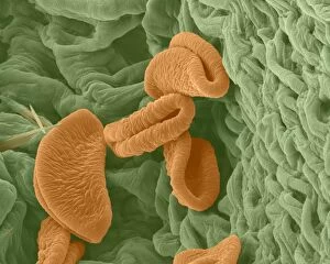
Scanning Electron micrograph (SEM): Open Stoma of a plant leaf; Date:

Background imageMicroscope Collection: Picture No. 10877013

Picture No. 10877013
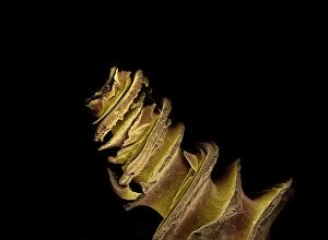
Scanning Electron Micrograph (SEM); Gecko Claw: Date:

Background imageMicroscope Collection: Picture No. 10877021

Picture No. 10877021
Scanning Electron micrograph (SEM): Open Stoma of a plant leaf; Date:

Background imageMicroscope Collection: Picture No. 10877014

Picture No. 10877014
Scanning Electron Micrograph (SEM): Gecko foot hairs Date:

Background imageMicroscope Collection: Picture No. 10877012

Picture No. 10877012
Scanning Electron Micrograph (SEM): human intestine showing villi Date:

Background imageMicroscope Collection: Picture No. 10877008

Picture No. 10877008
Scanning Electron Micrograph (SEM) of a resin cast of blood vessels in the intestines; Date:

Background imageMicroscope Collection: Picture No. 10877005

Picture No. 10877005
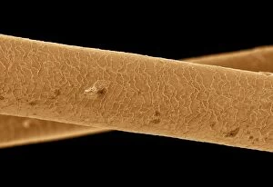
Scanning Electron Micrograph (SEM): Human hair - Asian Date:

Background imageMicroscope Collection: Picture No. 10877007

Picture No. 10877007
Scanning Electron Micrograph (SEM): Human Hairs Date:

Background imageMicroscope Collection: Picture No. 10876998

Picture No. 10876998
Scanning Electron Micrograph (SEM): Human Skin Date:

Background imageMicroscope Collection: Picture No. 10877000

Picture No. 10877000
Scanning Electron Micrograph (SEM): Human Skin Date:

Background imageMicroscope Collection: Picture No. 10877004

Picture No. 10877004
Scanning Electron Micrograph (SEM): Human hair - European Date:

Background imageMicroscope Collection: Picture No. 10877006

Picture No. 10877006
Scanning Electron Micrograph (SEM): Human pubic hair Date:

Background imageMicroscope Collection: Picture No. 10877003

Picture No. 10877003
Scanning Electron Micrograph (SEM): Human hair - African Date:


All Professionally Made to Order for Quick Shipping

"Unveiling the Unseen: Exploring the Microscopic World" Step into a realm of hidden wonders, where scientists like Rosalind Franklin and Santiago Ramon y Cajal have unraveled the mysteries through their microscopic lens. From Liver Fluke to Norovirus particles, these pioneers have paved the way for groundbreaking discoveries. Witness the intricate beauty of Snail teeth and Fat cells as they come alive under the microscope's gaze. Marvel at Lubbock's Graphic 84, a visual masterpiece capturing the essence of this microcosmic universe. Delve deeper into this microscopic world and encounter Plasmodium sp. , a malarial parasite that has plagued humanity for centuries. Explore E. Coli bacterium in all its glory, revealing both its potential dangers and scientific significance. Through microscope slide preparation techniques, we gain insight into complex organisms like never before. Discover how Fleming used his photographic skills to document crucial moments in microbiology history. The microscope acts as our window into an unseen dimension - it allows us to explore realms beyond our naked eye's reach, and is through this powerful tool that we continue to unravel nature's secrets one magnified image at a time. So join us on this journey "Under the Microscope, " where every slide holds a story waiting to be told – stories that shape our understanding of life itself.