Fraud Blocker Skip to main content

Microscope Collection (page 10)

"Unveiling the Unseen: Exploring the Microscopic World" Step into a realm of hidden wonders

Background imageMicroscope Collection: Suffragette Flora Drummond Stanhope Toy

Suffragette Flora Drummond Stanhope Toy
A Stanhope or optical toy, a device which allows the viewing of microphotographs without using a microscope. The stanhope was invented in 1857 by Rene Dagron, a French photographer
Background imageMicroscope Collection: Boy with microscope

Boy with microscope
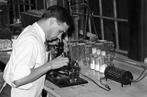
A boy in white overalls, sitting at a table examining something through a microscope. Date: 20th century

Background imageMicroscope Collection: Plague scientist in China

Plague scientist in China
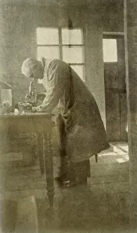
Western doctor or scientist in China during the bubonic plague outbreak; died during research work. Date: 1911

Background imageMicroscope Collection: Boys with Microscope

Boys with Microscope
Two schoolboys with a microscope in a classroom laboratory. Date: early 1970s

Background imageMicroscope Collection: Coffee under microscope

Coffee under microscope
Illustration of a coffee under microscope

Background imageMicroscope Collection: ALAN HODGKIN (1914-1998). English physiologist. Seated at microscope. Photograph, 1963

ALAN HODGKIN (1914-1998). English physiologist. Seated at microscope. Photograph, 1963

Background imageMicroscope Collection: JANE COOKE WRIGHT (1919-2013). American surgeon and cancer researcher. Photograph by Irwin Gooen

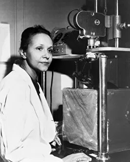
JANE COOKE WRIGHT (1919-2013). American surgeon and cancer researcher. Photograph by Irwin Gooen, 1958

Background imageMicroscope Collection: HOWARD UNIVERSITY, c1900. A class in the bacteriology laboratory at Howard University

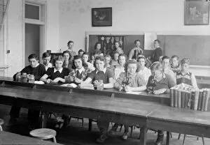
HOWARD UNIVERSITY, c1900. A class in the bacteriology laboratory at Howard University in Washington, D.C. Photograph, c1900

Background imageMicroscope Collection: MICROSCOPE, c1943. A woman using a microscope. Photograph, c1943

MICROSCOPE, c1943. A woman using a microscope. Photograph, c1943

Background imageMicroscope Collection: INFERTILITY CLINIC, 1956. Chief microbiologist Henry Isenberg examining a specimen

INFERTILITY CLINIC, 1956. Chief microbiologist Henry Isenberg examining a specimen at the Long Island Jewish Hospital Infertility Clinic in New Hyde Park, New York. Photograph, by Al Ravenna, 1956

Background imageMicroscope Collection: POLIOVIRUS, 1953. An early image of the poliovirus taken by an electron microscope

POLIOVIRUS, 1953. An early image of the poliovirus taken by an electron microscope. Photograph, 1953

Background imageMicroscope Collection: HIGH SCHOOL CLASS, c1936. Students in a science classroom at Montgomery High School

HIGH SCHOOL CLASS, c1936. Students in a science classroom at Montgomery High School in Montgomery County, Maryland, c1936

Background imageMicroscope Collection: Microscope

Microscope
Antique illustration of a microscope

Background imageMicroscope Collection: Compound microscope

Compound microscope
Antique illustration of a Compound microscope

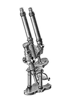
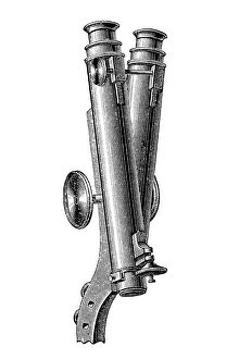
Background imageMicroscope Collection: Binocular Microscope

Binocular Microscope
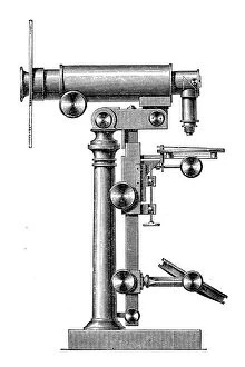
Antique illustration of a Binocular Microscope

Background imageMicroscope Collection: Microscopes

Microscopes
Antique illustration engraving of a various microscopes

Background imageMicroscope Collection: Compound microscope by Edmund Hartnack

Compound microscope by Edmund Hartnack
Antique illustration of a compound microscope made by Edmund Hartnack

Background imageMicroscope Collection: Binocularly microscope

Binocularly microscope
Antique illustration of a binocularly microscope

Background imageMicroscope Collection: Zeiss dissecting microscope

Zeiss dissecting microscope
Antique illustration of a Zeiss dissecting microscope

Background imageMicroscope Collection: Wenham Binocular Microscope

Wenham Binocular Microscope
Antique illustration of a WENHAM BINOCULAR MICROSCOPE

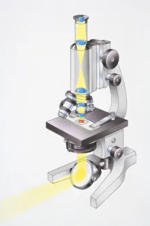
Background imageMicroscope Collection: Microscope, light passing through lenses and focusing on glass slide

Microscope, light passing through lenses and focusing on glass slide, light also reflecting from mirror onto underside of object being viewed, angled view

Background imageMicroscope Collection: Digital illustration of a microscope

Digital illustration of a microscope

Background imageMicroscope Collection: Digital illustration of scientist looking through microscope with woman standing behind

Digital illustration of scientist looking through microscope with woman standing behind

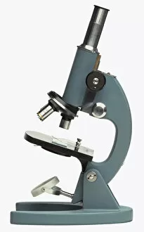
Background imageMicroscope Collection: Microscope

Microscope

Background imageMicroscope Collection: CHEMIST, 1938. Chemist running microscopic experiment on sweet potato starch, Laurel, Mississippi

CHEMIST, 1938. Chemist running microscopic experiment on sweet potato starch, Laurel, Mississippi. Photograph by Russell Lee, November 1938

Background imageMicroscope Collection: ROBERT ANDREWS MIILLIKAN (1868-1953). American physicist: oil over a photograph, c1930

ROBERT ANDREWS MIILLIKAN (1868-1953). American physicist: oil over a photograph, c1930

Background imageMicroscope Collection: Source Size = 1748 x 2519

Source Size = 1748 x 2519
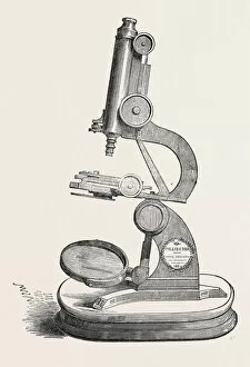
Pillischers Microscope, the Great Exhibition

Background imageMicroscope Collection: Granton Quarry, Near Edinburgh, Microscope Room and Library, Engraving 1884, Uk, Britain

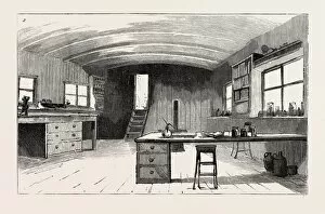
Granton Quarry, Near Edinburgh, Microscope Room and Library, Engraving 1884, Uk, Britain
GRANTON QUARRY, NEAR EDINBURGH, MICROSCOPE ROOM AND LIBRARY, engraving 1884, UK, britain, british, europe, united kingdom, great britain, european

Background imageMicroscope Collection: Advertisement for Cadburys Cocoa, from Nature, 20th November 1890

Advertisement for Cadburys Cocoa, from Nature, 20th November 1890
XJF270048 Advertisement for Cadburys Cocoa, from Nature, 20th November 1890 (litho) (b/w photo) (litho) (b/w photo) by English School, (19th century) (after); Private Collection; English

Background imageMicroscope Collection: Portrait of Alphonse Leroy, 1783 (oil on canvas)

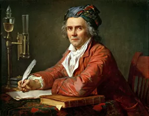
Portrait of Alphonse Leroy, 1783 (oil on canvas)
XIR212875 Portrait of Alphonse Leroy, 1783 (oil on canvas) by David, Jacques Louis (1748-1825); 72x91 cm; Musee Fabre, Montpellier, France; French, out of copyright

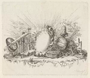
Background imageMicroscope Collection: Cartouche surrounded by scientific instruments, Johannes Petrus van Horstok, 1755 - 1825

Cartouche surrounded by scientific instruments, Johannes Petrus van Horstok, 1755 - 1825

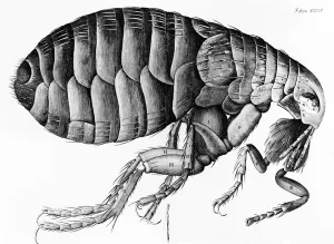
Background imageMicroscope Collection: A Flea from Microscope Observation by Robert Hooke (1635-1703), 1665 (engraving)

A Flea from Microscope Observation by Robert Hooke (1635-1703), 1665 (engraving)
XJF131255 A Flea from Microscope Observation by Robert Hooke (1635-1703), 1665 (engraving) (b/w photo) by English School, (17th century); Private Collection; English, out of copyright

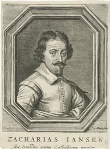
Background imageMicroscope Collection: Portrait of Zacharias Jansen, 1655 (engraving)

Portrait of Zacharias Jansen, 1655 (engraving)
XOS1766231 Portrait of Zacharias Jansen, 1655 (engraving) by Meurs, Jacob van (c.1619-a.1680); 18.8x14 cm; Rijksmuseum, Amsterdam

Background imageMicroscope Collection: Compound Microscope and Case

Compound Microscope and Case; Micrometric stage invented by Michel-Ferdinand d Albert d Ailly, sixth duke of Chaulnes, French, 1714 - 1769, Gilt-bronze mounts attributed to Jacques Caffieri, French

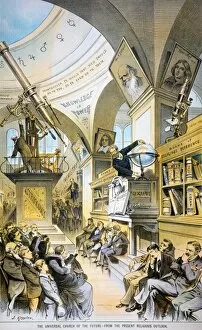
Background imageMicroscope Collection: RELIGIOUS OUTLOOK, 1883. The Universal Church of the Future-From the Present Religious Outlook

RELIGIOUS OUTLOOK, 1883. The Universal Church of the Future-From the Present Religious Outlook. Lithograph cartoon by Joseph Keppler, 1883

Background imageMicroscope Collection: HOOKEs MICROGRAPHIA. Title-page of the first edition of Robert Hookes Micrographia

HOOKEs MICROGRAPHIA. Title-page of the first edition of Robert Hookes Micrographia, London, 1665, an early landmark in microscopy containing the first illustration of cells

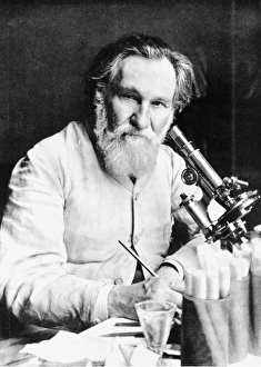
Background imageMicroscope Collection: ILYA ILICH MECHNIKOV (1845-1916). Russian bacteriologist and zoologist

ILYA ILICH MECHNIKOV (1845-1916). Russian bacteriologist and zoologist

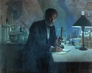
Background imageMicroscope Collection: LOUIS PASTEUR (1822-1895). French chemist and microbiologist. Pasteur in his laboratory

LOUIS PASTEUR (1822-1895). French chemist and microbiologist. Pasteur in his laboratory. Fresco by Fournier

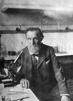
Background imageMicroscope Collection: ELIE METCHNIKOFF (1845-1916). Russian zoologist and bacteriologist

ELIE METCHNIKOFF (1845-1916). Russian zoologist and bacteriologist

Background imageMicroscope Collection: Male doctor using microscope in surgery, (B&W)

Male doctor using microscope in surgery, (B&W)

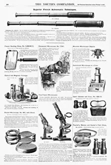
Background imageMicroscope Collection: SCIENTIFIC INSTRUMENTS, 1890. American magazine advertisements for various scientific instruments

SCIENTIFIC INSTRUMENTS, 1890. American magazine advertisements for various scientific instruments, 1890

Background imageMicroscope Collection: Robert Hookes Microscope

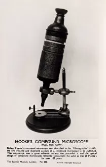
Robert Hookes Microscope
Full-size copy of Robert Hookes Compound Microscope - held at the Science Museum, London. Hooke, an English natural philosopher, architect and polymath (1635-1703) - author of Micrographia (1665)

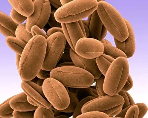
Background imageMicroscope Collection: Picture No. 11014617

Picture No. 11014617
Veronica Pollen Scanning Electron Micrograph (SEM): Magnification x8220 (if print A4 size: 29.7 cm wide) - These small pollen grains are sticky and are carried away by pollinating insects Date:

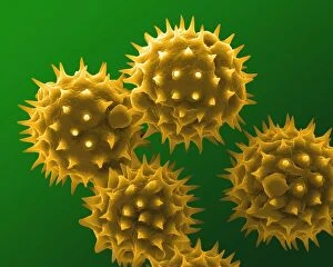
Background imageMicroscope Collection: Picture No. 11014615

Picture No. 11014615
Sunflower Pollen Scanning Electron Micrograph (SEM): Magnification x10, 000 (if print A4 size: 29.7 cm wide) - The large flowers are insect pollinated

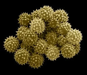
Background imageMicroscope Collection: Picture No. 11014607

Picture No. 11014607
Groundsel Pollen Scanning Electron Micrograph (SEM): Magnification 2 x6940 (if print A4 size: 29.7 cm wide) - Groundsel is a common weed - It is insect pollinated; provides food for many insects


All Professionally Made to Order for Quick Shipping

"Unveiling the Unseen: Exploring the Microscopic World" Step into a realm of hidden wonders, where scientists like Rosalind Franklin and Santiago Ramon y Cajal have unraveled the mysteries through their microscopic lens. From Liver Fluke to Norovirus particles, these pioneers have paved the way for groundbreaking discoveries. Witness the intricate beauty of Snail teeth and Fat cells as they come alive under the microscope's gaze. Marvel at Lubbock's Graphic 84, a visual masterpiece capturing the essence of this microcosmic universe. Delve deeper into this microscopic world and encounter Plasmodium sp. , a malarial parasite that has plagued humanity for centuries. Explore E. Coli bacterium in all its glory, revealing both its potential dangers and scientific significance. Through microscope slide preparation techniques, we gain insight into complex organisms like never before. Discover how Fleming used his photographic skills to document crucial moments in microbiology history. The microscope acts as our window into an unseen dimension - it allows us to explore realms beyond our naked eye's reach, and is through this powerful tool that we continue to unravel nature's secrets one magnified image at a time. So join us on this journey "Under the Microscope, " where every slide holds a story waiting to be told – stories that shape our understanding of life itself.