Fraud Blocker Skip to main content

Microscopic Photos Collection (page 6)

Discovering the hidden wonders of the microscopic world is like entering a realm of infinite fascination

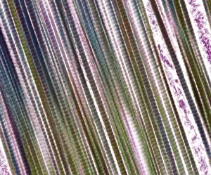
Background imageMicroscopic Photos Collection: Tendon fibres, TEM

Tendon fibres, TEM
Tendon fibres, coloured transmission electron micrograph (TEM). A tendon is made up of parallel bundles of collagen fibres

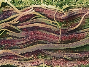
Background imageMicroscopic Photos Collection: Coloured SEM of skeletal (striated) muscle fibres

Coloured SEM of skeletal (striated) muscle fibres
Skeletal muscle. Coloured scanning electron micrograph (SEM) of skeletal muscle fibres. Muscle fibres are extremely long, cylindrical cells

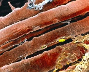
Background imageMicroscopic Photos Collection: Coloured SEM of a bundle of skeletal muscle fibres

Coloured SEM of a bundle of skeletal muscle fibres

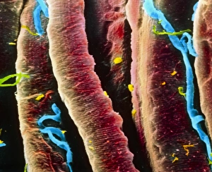
Background imageMicroscopic Photos Collection: False-colour SEM of skeletal muscle fibres

False-colour SEM of skeletal muscle fibres
Muscle. False-colour scanning electron micrograph of a bundle of skeletal muscle fibres (red), also known as striated muscle fibres, and a few capillaries (blue)

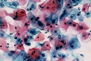
Background imageMicroscopic Photos Collection: LM of cervical smear cells with mild dyskaryosis

LM of cervical smear cells with mild dyskaryosis

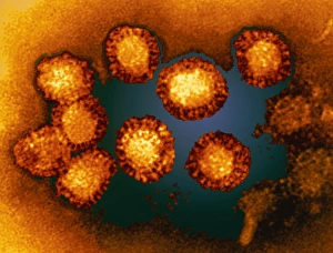
Background imageMicroscopic Photos Collection: West Nile viruses

West Nile viruses. Coloured transmission electron micrograph (TEM) of a group of West Nile viruses (WNV). This virus is known to cause encephalitis in humans

Background imageMicroscopic Photos Collection: Computer artwork of osteoporosis affecting bone

Computer artwork of osteoporosis affecting bone
Osteoporosis. Computer artwork of a Scanning Electron Micrograph showing the effect of osteoporosis on bone tissue. This section through one of the vertebrae of the backbone shows the cancellous

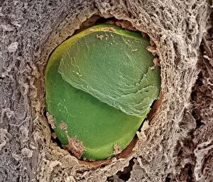
Background imageMicroscopic Photos Collection: Embolism

Embolism. Coloured scanning electron micrograph (SEM) of a blockage, or embolism, in a blood vessel. Embolisms, particularly when they occur in vital organs such as the lungs (pulmonary embolism)

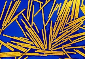
Background imageMicroscopic Photos Collection: Coloured TEM of Tobacco Mosaic Viruses (TMV)

Coloured TEM of Tobacco Mosaic Viruses (TMV)
Tobacco mosaic viruses. Coloured transmission electron micrograph of a cluster of rod-shaped red virus particles of tobacco mosaic virus (TMV)

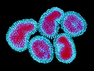
Background imageMicroscopic Photos Collection: Influenza viruses

Influenza viruses. Coloured transmission electron micrograph (TEM) of influenza (flu) viruses. The flu virus belongs to the orthomyxovirus group of viruses which have an affinity for mucus

Background imageMicroscopic Photos Collection: Coloured TEM of HHV6 infecting a cell

Coloured TEM of HHV6 infecting a cell
Coloured transmission electron micrograph of Human Herpes Virus type 6 (HHV6) infecting a human cell. Within a pseudopodium (cell outgrowth centre)

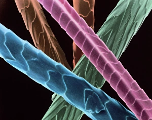
Background imageMicroscopic Photos Collection: Angora wool

Angora wool fibres. Coloured scanning electron micrograph of fibres of angora wool. These show the overlapping scales typical of animal hairs

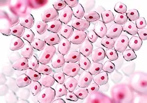
Background imageMicroscopic Photos Collection: Cells

Cells. Computer artwork of non-specific cells. The red dot in the centre of each cell is the nucleus where the DNA (deoxyribonucleic acid) of each cell is stored

Background imageMicroscopic Photos Collection: Stressed culture cell, SEM

Stressed culture cell, SEM

Background imageMicroscopic Photos Collection: Pancreas cell, SEM

Pancreas cell, SEM
Pancreas cell. Coloured scanning electron micrograph (SEM) of an acinar (exocrine) pancreatic cell. Acinar cells produce and excrete digestive enzymes to the small intestine

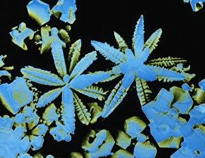
Background imageMicroscopic Photos Collection: SEM of snow & ice crystals

SEM of snow & ice crystals
False-colour scanning electron micrograph of a pair of tiny, flower-like ice crystals. Snow & ice crystals have a characteristic hexagonal symmetry

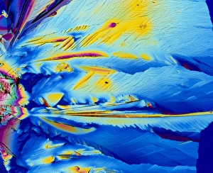
Background imageMicroscopic Photos Collection: Oestradiol, female sex hormone

Oestradiol, female sex hormone
Female sex hormone. Polarised light micrograph of crystals of Oestradiol, also known as beta- Estradiol. This is the most potent of the six naturally-occurring oestrogens

Background imageMicroscopic Photos Collection: Serotonin crystals, light micrograph

Serotonin crystals, light micrograph
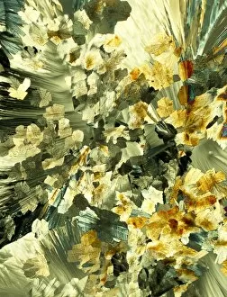
Serotonin. Polarised light micrograph of serotonin, a neurotransmitter. Serotonin is derived from tryptophan, and is found in the vertebrate brain

Background imageMicroscopic Photos Collection: Syenite, thin section, polarized LM

Syenite, thin section, polarized LM
Polarised light micrograph of a thin section of syenite, a coarse-grained igneous rock, containing the mineral augite. Augite occurs as a prismatic crystal which in cross-section appears almost

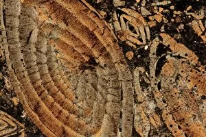
Background imageMicroscopic Photos Collection: Limestone, thin section, polarised LM

Limestone, thin section, polarised LM
Nummulitic limestone. Polarised light micrograph of a thin section of a Nummulitic limestone. The large coloured objects are the remains of large foraminifera, Nummulites sp

Background imageMicroscopic Photos Collection: Quartzite, thin section, polarized LM

Quartzite, thin section, polarized LM
Polarised light micrograph of a thin section of quartzite, a metamorphic rock. The composition of quartzite varies: quartz is an essential component, with accessories of mica and feldspar

Background imageMicroscopic Photos Collection: Salt crystals, light micrograph

Salt crystals, light micrograph
Salt. Dark field light micrograph of sodium chloride crystals, the common salt. This is a white crystalline compound widespread in nature as a solid (rock salt)

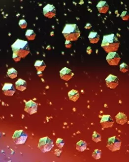
Background imageMicroscopic Photos Collection: Insulin crystals, light micrograph

Insulin crystals, light micrograph
Insulin. Polarised light micrograph (PLM) of crystals of the hormone insulin. The crystals appear hexagonal in shape. The insulin molecule is made up of two chains of amino acids (A & B chains)

Background imageMicroscopic Photos Collection: Purkinje nerve cells, light micrograph

Purkinje nerve cells, light micrograph
Purkinje nerve cells. Light micrograph of a section through a row of Purkinje nerve cells from the brains cerebellum. Each Purkinje cell is composed of a flask-shaped cell body which branch numerous

Background imageMicroscopic Photos Collection: Silver Nitrate, light micrograph

Silver Nitrate, light micrograph
Composite crystals of Silver Nitrate, polarised light micrograph

Background imageMicroscopic Photos Collection: Euphorbia flower reproductive parts, SEM

Euphorbia flower reproductive parts, SEM
Euphorbia flower reproductive parts. Coloured scanning electron micrograph (SEM) of the reproductive parts of a Euphorbia supina flower

Background imageMicroscopic Photos Collection: Polarised LM of vitamin D crystals

Polarised LM of vitamin D crystals
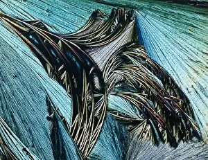
Vitamin D. Polarised light micrograph of crystals of vitamin D. Vitamin D is fat-soluble, and is found in foods such as animal fats, milk, butter and eggs

Background imageMicroscopic Photos Collection: Polarised LM of niacinamide crystals

Polarised LM of niacinamide crystals
Nicotinamide. Polarised light micrograph of crystals of nicotinamide, or niacinamide. This is the amide of nicotinic acid. The acid and amide are both B vitamins, and are equally active

Background imageMicroscopic Photos Collection: Treponema pallidum bacteria

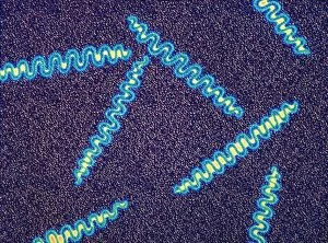
Treponema pallidum bacteria

Background imageMicroscopic Photos Collection: Gas gangrene bacteria, SEM

Gas gangrene bacteria, SEM
Gas gangrene bacteria (Clostridium perfringens), coloured scanning electron micrograph (SEM). These rod-shaped bacteria are pathogenic

Background imageMicroscopic Photos Collection: Tuberculosis bacteria, TEM

Tuberculosis bacteria, TEM
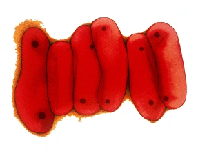
Tuberculosis bacteria. Coloured transmission electron micrograph (TEM) of a section through Mycobacterium tuberculosis bacteria

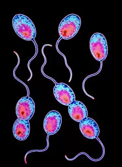
Background imageMicroscopic Photos Collection: Moraxella catarrhalis bacteria

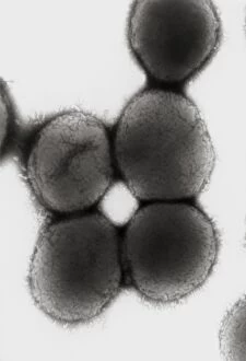
Moraxella catarrhalis bacteria

Background imageMicroscopic Photos Collection: Rhodopseudomonas sp bacteria

Rhodopseudomonas sp bacteria

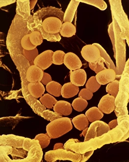
Background imageMicroscopic Photos Collection: SEM of soil bacteria

SEM of soil bacteria

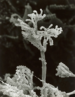
Background imageMicroscopic Photos Collection: Penicillin fungus growing on cheddar cheese

Penicillin fungus growing on cheddar cheese
Scanning electron micrograph (SEM) the fruiting bodies or conidia of a penicillin fungus growing on a piece of English cheddar cheese which has gone mouldy in the refrigerator

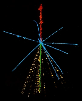
Background imageMicroscopic Photos Collection: Emulsion photo of cosmic ray event

Emulsion photo of cosmic ray event
The power of a cosmic ray is shown in this false- colour emulsion photo of a cosmic ray sulphur nucleus (red) colliding with a nucleus in the emulsion

Background imageMicroscopic Photos Collection: Particle tracks on a star chart

Particle tracks on a star chart

Background imageMicroscopic Photos Collection: Disc floret, SEM

Disc floret, SEM
Disc floret. Coloured scanning electron micrograph (SEM) of a disc floret of a composite flower. Daisies (family Asteraceae, formerly Compositae) have inflorescences composed of two types of flowers

Background imageMicroscopic Photos Collection: Comfrey flower petal, SEM

Comfrey flower petal, SEM
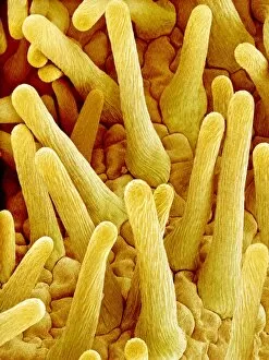
Comfrey flower petal. Coloured scanning electron micrograph (SEM) showing papillae on a petal from a comfrey (Symphytum officinale) flower. Papillae are projections from epidermal cells


All Professionally Made to Order for Quick Shipping

Discovering the hidden wonders of the microscopic world is like entering a realm of infinite fascination. From particle tracks leaving trails of mystery to the mesmerizing bubble chamber photo capturing the decay of a sigma particle, these microscopic photos unveil secrets that lie beyond our naked eye's reach. One remarkable image showcases the first observation of an omega-minus particle, a groundbreaking moment in scientific history. Meanwhile, another snapshot reveals the delicate beauty of a human blastocyst, reminding us of life's miraculous beginnings. Immunofluorescent LM offers us glimpses into intricate networks within our own bodies - neurons and astrocytes intricately woven together like cosmic constellations. And then there are oxytocin hormone crystals, their vibrant colors shining through as if encapsulating love itself. The iris of an eye captured by SEM reminds us that even something as ordinary as our eyes holds extraordinary complexity when seen up close. Similarly, osteoporotic bone under SEM unveils its fragile structure with astonishing detail. Light micrographs bring forth stunning images such as oxytocin crystals glistening like precious gems or spiral spore chains formed by Streptomyces bacteria resembling ethereal works of art. And let us not forget about our retina - a gateway to vision and perception - or nerve cells firing messages at lightning speed throughout our bodies, orchestrating every movement and thought we have. These microscopic photos offer more than just visual delight; they invite us to ponder upon the vastness contained within each minuscule frame. They remind us that sometimes it is in exploring the smallest details that we gain profound insights into ourselves and the world around us.