
Molecular Collection (page 2)
"Molecular Marvels: Unveiling the Intricate World of Rosalind Franklin and DNA" Delving into the realm wonders, we encounter the brilliant mind of Rosalind Franklin


All Professionally Made to Order for Quick Shipping
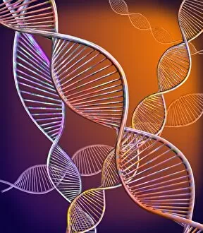
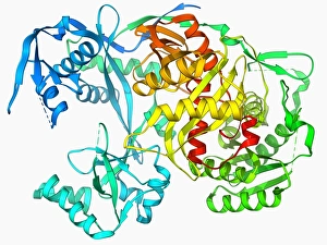
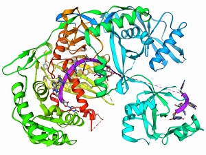
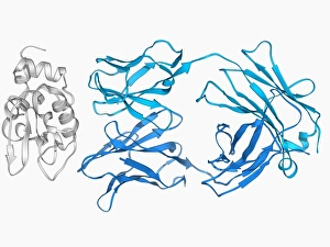
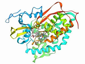
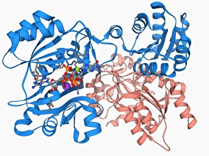
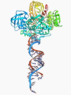
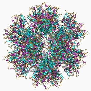
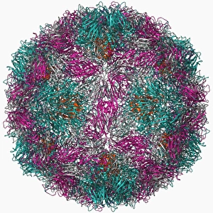
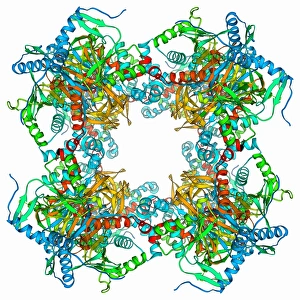
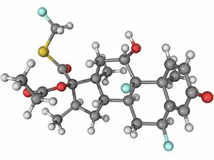
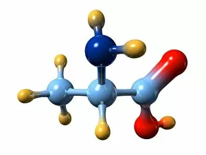
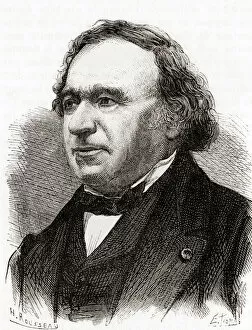
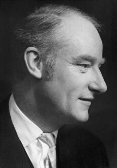
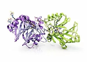
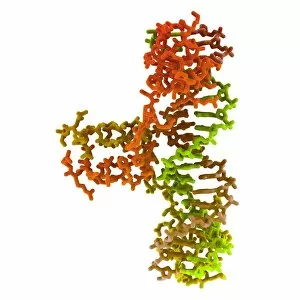
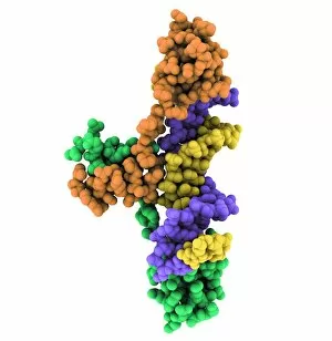
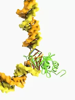
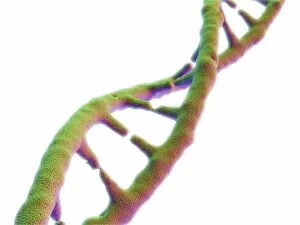
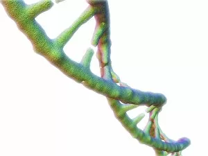
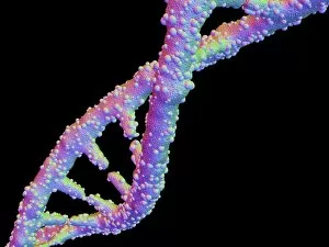
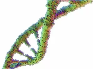
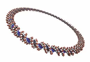
"Molecular Marvels: Unveiling the Intricate World of Rosalind Franklin and DNA" Delving into the realm wonders, we encounter the brilliant mind of Rosalind Franklin, whose groundbreaking work paved the way for our understanding of life's blueprint. Her pioneering research on X-ray crystallography revealed a mesmerizing image – the double-stranded RNA molecule, unraveling nature's secrets strand by strand. Intriguingly intricate, DNA transcription comes to life as we explore its molecular model, and is through this process that genetic information is transcribed from DNA to RNA, orchestrating the symphony of life itself. James Clerk Maxwell's caricature reminds us of his profound contributions to electromagnetism and how it laid the foundation for comprehending molecular interactions at an atomic level. His genius echoes through time as we marvel at his caricatured presence. Shifting gears towards medicinal breakthroughs, let us not overlook Amitriptyline antidepressant molecule – a tiny compound with enormous potential in alleviating human suffering. Its structure represents hope and relief for those battling mental health challenges. Art meets science when we encounter metabolic enzyme artwork; a visual representation showcasing these powerful catalysts that drive countless biochemical reactions within our bodies. Their elegant complexity highlights their indispensable role in sustaining life's delicate balance. Computer-generated models bring forth a vivid depiction of DNA molecules – intricate helices intertwining like cosmic dancers choreographed by evolution itself. These virtual representations invite us to delve deeper into their mysteries while appreciating their breathtaking beauty. The nucleosome molecule takes center stage as it reveals how DNA wraps around histone proteins forming chromatin structures within our cells' nuclei. This architectural masterpiece ensures proper gene regulation and compaction while offering glimpses into cellular harmony on a microscopic scale. Abstract images portraying DNA molecules captivate our imagination with vibrant colors and patterns reminiscent of unseen universes hidden within each cell nucleus—a testament to nature's artistic prowess.