Fraud Blocker Skip to main content

Molecular Collection (page 9)

"Molecular Marvels: Unveiling the Intricate World of Rosalind Franklin and DNA" Delving into the realm wonders, we encounter the brilliant mind of Rosalind Franklin

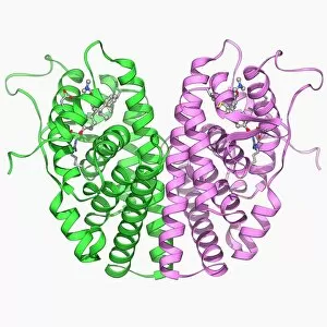
Background imageMolecular Collection: Oestrogen receptor and tamoxifen drug F006 / 9674

Oestrogen receptor and tamoxifen drug F006 / 9674
Oestrogen receptor and tamoxifen drug, molecular model. Tamoxifen is a selective oestrogen receptor modulator (SERM) that blocks oestrogen production in breast tissue by inhibiting the oestrogen

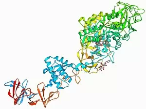
Background imageMolecular Collection: Glucansucrase enzyme molecule F006 / 9685

Glucansucrase enzyme molecule F006 / 9685
Glucansucrase, molecular model. This enzyme catalyses the cleavage of sucrose into fructose and glucose

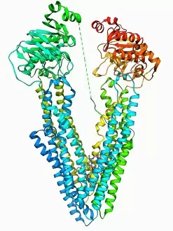
Background imageMolecular Collection: Cytochrome BC1 molecule F006 / 9681

Cytochrome BC1 molecule F006 / 9681
Cytochrome BC1, Molecular model. Cytochrome molecules perform oxidation and reduction reactions for electron transport, a chain of reactions used to power cellular processes that require energy

Background imageMolecular Collection: HIV reverse transcription enzyme F006 / 9684

HIV reverse transcription enzyme F006 / 9684
HIV reverse transcription enzyme. Molecular model of the reverse transcriptase enzyme found in HIV (the human immunodeficiency virus) bound to the inhibitor nevirapine

Background imageMolecular Collection: Human growth hormone molecule F006 / 9683

Human growth hormone molecule F006 / 9683
Human growth hormone. Molecular model of human growth hormone (hGH, orange) bound to the extracellular domain of the human growth hormone binding protein (hGHBP)

Background imageMolecular Collection: HIV DNA and transcription factor F006 / 9680

HIV DNA and transcription factor F006 / 9680
HIV DNA and transcription factor. Molecular model of DNA (deoxyribonucleic acid) from HIV-1 (human immunodeficiency virus type 1) complexed with the transcription factor kappa B

Background imageMolecular Collection: Self-assembled DNA triangle F006 / 9679

Self-assembled DNA triangle F006 / 9679
Self-assembled DNA triangle. Molecular model of DNA (deoxyribonucleic acid) strands forming what is called a tensegrity triangle

Background imageMolecular Collection: Human pathogen recognition molecule F006 / 9677

Human pathogen recognition molecule F006 / 9677
Human pathogen recognition molecule. Molecular model of a complex between human lymphocyte antigen 96 and toll-like receptor 4

Background imageMolecular Collection: Antibodies and their antigen F006 / 9678

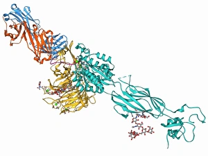
Antibodies and their antigen F006 / 9678
Antibodies and their antigen. Molecular model showing the molecular structure of two anti-P-glycoprotein antibodies (upper left and right) and the P-glycoprotein (v-shaped) to which they respond

Background imageMolecular Collection: Bacterial nanocompartment F006 / 9672

Bacterial nanocompartment F006 / 9672
Bacterial nanocompartment. Molecular molecule of a nanocompartment from the bacterium Thermotoga maritima. This is a shell formed from encapsulin proteins that encloses enzymes that defend the cell

Background imageMolecular Collection: E coli virulence factor molecule F006 / 9675

E coli virulence factor molecule F006 / 9675
E. coli virulence factor molecule. Molecular model of the enzyme arylsulfate sulfotransferase (ASST) from an Escherichia coli bacterium

Background imageMolecular Collection: Fatty acid binding protein and inhibitor F006 / 9676

Fatty acid binding protein and inhibitor F006 / 9676
Fatty acid binding protein and inhibitor. Molecular model of adipocyte fatty-acid binding protein (A-FABP) bound to an inhibitor

Background imageMolecular Collection: Fenna-Matthews-Olson complex molecule F006 / 9673

Fenna-Matthews-Olson complex molecule F006 / 9673
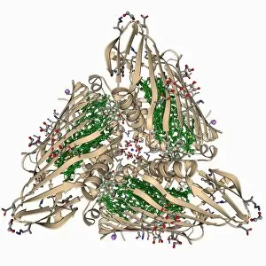
Fenna-Matthews-Olson complex. Molecular model of the Fenna-Matthews-Olson complex from the green sulphur bacterium Prosthecochloris aestuarii

Background imageMolecular Collection: Nucleoside diphosphate kinase molecule F006 / 9671

Nucleoside diphosphate kinase molecule F006 / 9671
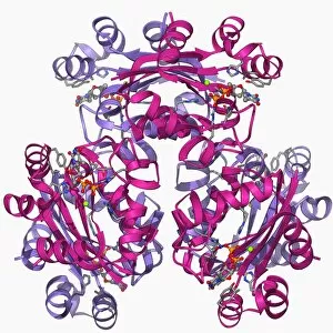
Nucleoside diphosphate kinase. Molecular model of the enzyme nucleoside diphosphate kinase from the Acanthamoeba polyphaga mimivirus

Background imageMolecular Collection: Cytochrome C, molecular model F006 / 9670

Cytochrome C, molecular model F006 / 9670
Cytochrome C, molecular model. Cytochrome molecules perform oxidation and reduction reactions for electron transport, a chain of reactions used to power cellular processes that require energy

Background imageMolecular Collection: Concanavalin A F006 / 9668

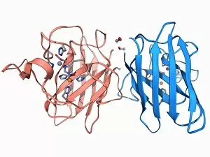
Concanavalin A F006 / 9668
Concanavalin A, molecular model. This lectin protein is found in legumes. Lectins are highly specific sugar-binding proteins that play a role in biological recognition

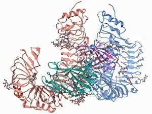
Background imageMolecular Collection: Toll-like receptor 3 and RNA F006 / 9666

Toll-like receptor 3 and RNA F006 / 9666
Toll-like receptor 3 and RNA. Molecular model of the toll-like receptor 3 (TLR3) protein (pink and blue) bound to a strand of RNA (ribonucleic acid, green and yellow)

Background imageMolecular Collection: Calcium-binding protein molecule F006 / 9667

Calcium-binding protein molecule F006 / 9667
Calcium-binding protein. Molecule model of the calcium-binding protein calmodulin (CaM). This protein is found in all eukaryotic cells

Background imageMolecular Collection: Isocitrate dehydrogenase molecule F006 / 9663

Isocitrate dehydrogenase molecule F006 / 9663
Isocitrate dehydrogenase, molecular model. This enzyme catalyses the third step in the citric acid (or Krebs) cycle, the process by which mitochondria convert glucose to energy

Background imageMolecular Collection: Opsin molecule F006 / 9665

Opsin molecule F006 / 9665
Opsin. Molecular model of a ligand-free opsin molecule. Opsins are found in photoreceptor cells (rods and cones) in the retina of the eye

Background imageMolecular Collection: Interferon antagonism by viral protein F006 / 9662

Interferon antagonism by viral protein F006 / 9662
Interferon (IFN) antagonism by viral protein. Molecular model of an orthopoxvirus IFN-gamma-binding protein bound to an IFN-gamma molecule

Background imageMolecular Collection: DNA and MECP2 complex, molecular model F006 / 9664

DNA and MECP2 complex, molecular model F006 / 9664
DNA and MECP2 complex. Molecular model of MECP2 (methyl CpG binding protein 2 (Rett syndrome)) bound to the BDNF (brain-derived neurotrophic factor)

Background imageMolecular Collection: Selenocysteine synthase enzyme molecule F006 / 9661

Selenocysteine synthase enzyme molecule F006 / 9661
Selenocysteine synthase enzyme molecule. Computer model showing the molecular structure of the enzyme selenocysteine synthase (SecS)

Background imageMolecular Collection: Androgen receptor and modulator F006 / 9660

Androgen receptor and modulator F006 / 9660
Androgen receptor and modulator. Molecular model of a selective androgen receptor modulator (SARM) bound to an androgen receptor

Background imageMolecular Collection: Bacterial biofilm enzyme F006 / 9658

Bacterial biofilm enzyme F006 / 9658
Bacterial biofilm enzyme. Molecular model of the enzyme glucansucrase from the bacterium Streptococcus mutans. This enzyme is used to form bacterial biofilms on teeth that cause dental caries

Background imageMolecular Collection: Rous sarcoma virus capsid F006 / 9653

Rous sarcoma virus capsid F006 / 9653
Rous sarcoma virus capsid, molecular model. In viruses, the capsid is the protein shell that encloses the genetic material

Background imageMolecular Collection: Rabbit eye lens protein molecule F006 / 9657

Rabbit eye lens protein molecule F006 / 9657
Rabbit eye lens protein. Molecular model of lambda-crystallin, a structural protein found in the eye lenses of rabbits (family Leporidae)

Background imageMolecular Collection: Sodium-potassium ion pump protein F006 / 9656

Sodium-potassium ion pump protein F006 / 9656
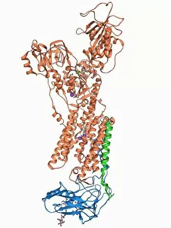
Sodium-potassium ion pump protein, molecular model. Sodium-potassium ATPase (adenosine triphosphatase) is an ATP-powered ion pump found in all animal cells

Background imageMolecular Collection: Chromatin remodelling factor and DNA F006 / 9655

Chromatin remodelling factor and DNA F006 / 9655
Chromatin remodelling factor and DNA, molecular model. The strands of DNA (deoxyribonucleic acid) are at left and right. This chromatin remodelling factor is ISW1a

Background imageMolecular Collection: 70S ribosome, molecular model F006 / 9651

70S ribosome, molecular model F006 / 9651
70S ribosome, molecular model. Ribosomes are composed of protein and RNA (ribonucleic acid). In bacteria each ribosome consists of a small (30S) subunit and a large (50S) subunit

Background imageMolecular Collection: MscS ion channel protein structure F006 / 9650

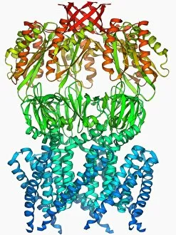
MscS ion channel protein structure F006 / 9650
MscS ion channel protein structure. Molecular model of a mechanosensitive channel of small conductance (MscS) from an Escherichia coli bacterium

Background imageMolecular Collection: Streptavidin bacterial protein F006 / 9654

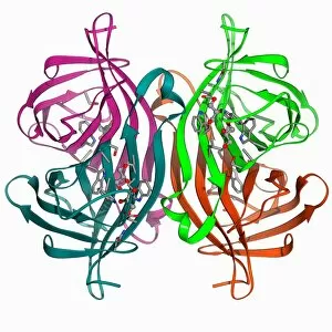
Streptavidin bacterial protein F006 / 9654
Streptavidin bacterial protein, molecular model. Streptavidin is a protein obtained from the bacterium Streptomyces avidinii

Background imageMolecular Collection: Fatty acid synthase molecule F006 / 9647

Fatty acid synthase molecule F006 / 9647
Fatty acid synthase (FAS), molecular model. FAS is a multi-enzyme that plays a key role in the synthesis of fatty acids (lipids) in the human body. It is not a single enzyme but a whole enzyme system

Background imageMolecular Collection: Eye lens protein molecule F006 / 9652

Eye lens protein molecule F006 / 9652
Eye lens protein. Molecular model of alpha-crystallin, a protein found in the lens of the eye. The regular arrangement of the protein in the lens is thought to be responsible for its transparency

Background imageMolecular Collection: Integrin and fibrinogen complex molecule F006 / 9649

Integrin and fibrinogen complex molecule F006 / 9649
Integrin and fibrinogen complex. Molecular model of integrin alpha-II beta-3 complexed with fibrinogen. Integrin alpha-II beta-3 is a transmembrane protein found on platelets

Background imageMolecular Collection: SelB elongation factor bound to RNA F006 / 9648

SelB elongation factor bound to RNA F006 / 9648
SelB elongation factor bound to RNA. Molecular model of the SelB elongation factor bound to an mRNA (messenger ribonucleic acid) hairpin formed by the selenocysteine insertion sequence (SECIS)

Background imageMolecular Collection: Voltage-gated potassium channel F006 / 9642

Voltage-gated potassium channel F006 / 9642
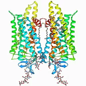
Voltage-gated potassium channel. Molecular model of a voltage-gated potassium (Kv) ion channel. Ion channels are membrane-spanning proteins that form pores in cell membranes

Background imageMolecular Collection: Src protein molecule F006 / 9646

Src protein molecule F006 / 9646
Src protein, molecular model. Src is a tyrosine kinase, a signalling protein in cells that has the ability to turn on protein synthesis and cellular growth

Background imageMolecular Collection: Copper, zinc superoxide dismutase enzyme F006 / 9645

Copper, zinc superoxide dismutase enzyme F006 / 9645
Copper, zinc superoxide dismutase enzyme

Background imageMolecular Collection: Fungal prion protein F006 / 9644

Fungal prion protein F006 / 9644
Fungal prion protein. Molecular model of the amyloid form of the HET prion protein

Background imageMolecular Collection: Beta-2 adrenergic receptor molecule F006 / 9643

Beta-2 adrenergic receptor molecule F006 / 9643
Beta-2 adrenergic receptor. Molecular model of a human beta-2 adrenergic receptor bound to an antibody. Beta receptors respond to adrenalin, causing a sympathetic (fight or flight) response

Background imageMolecular Collection: Adenovirus protein and tumor suppressor F006 / 9641

Adenovirus protein and tumor suppressor F006 / 9641
Adenovirus protein and tumour suppressor. Molecular model of the E1A protein from human adenovirus bound to a retinoblastoma tumour suppressor

Background imageMolecular Collection: Beta-2 adrenergic receptor molecule F006 / 9640

Beta-2 adrenergic receptor molecule F006 / 9640
Beta-2 adrenergic receptor. Molecular model of a human beta-2 adrenergic receptor bound to an antibody. Beta receptors respond to adrenalin, causing a sympathetic (fight or flight) response

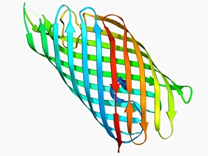
Background imageMolecular Collection: E coli virulence factor F006 / 9639

E coli virulence factor F006 / 9639
E. coli virulence factor. Molecular model of the beta-domain of the EspP autotransporter protein from the bacterium Escherichia coli

Background imageMolecular Collection: 70S ribosome, molecular model F006 / 9638

70S ribosome, molecular model F006 / 9638
70S ribosome. Molecular model of a 70S ribosome complex containing a Shine-Dalgarno helix, the point of mRNA (messenger ribonucleic acid) binding

Background imageMolecular Collection: Metal-sensing RNA molecule F006 / 9636

Metal-sensing RNA molecule F006 / 9636
Metal-sensing RNA molecule. Molecular model of an M-box riboswitch, a length of RNA (ribonucleic acid) that regulates levels of metal ions in a cell

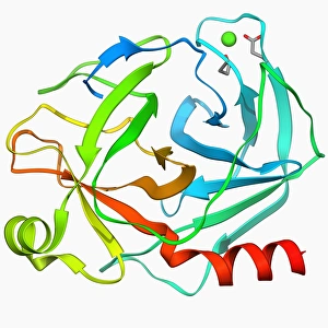
Background imageMolecular Collection: Trypsin molecule F006 / 9634

Trypsin molecule F006 / 9634
Trypsin molecule. Molecular model of the digestive protease enzyme trypsin. Trypsin is released by the pancreas to break down proteins into smaller chains of amino acids

Background imageMolecular Collection: PolyA polymerase and RNA F006 / 9635

PolyA polymerase and RNA F006 / 9635
Poly(A) polymerase and RNA. Molecular model of poly(A) polymerase complexed with RNA (ribonucleic acid) and ATP (adenosine triphosphate)


All Professionally Made to Order for Quick Shipping

"Molecular Marvels: Unveiling the Intricate World of Rosalind Franklin and DNA" Delving into the realm wonders, we encounter the brilliant mind of Rosalind Franklin, whose groundbreaking work paved the way for our understanding of life's blueprint. Her pioneering research on X-ray crystallography revealed a mesmerizing image – the double-stranded RNA molecule, unraveling nature's secrets strand by strand. Intriguingly intricate, DNA transcription comes to life as we explore its molecular model, and is through this process that genetic information is transcribed from DNA to RNA, orchestrating the symphony of life itself. James Clerk Maxwell's caricature reminds us of his profound contributions to electromagnetism and how it laid the foundation for comprehending molecular interactions at an atomic level. His genius echoes through time as we marvel at his caricatured presence. Shifting gears towards medicinal breakthroughs, let us not overlook Amitriptyline antidepressant molecule – a tiny compound with enormous potential in alleviating human suffering. Its structure represents hope and relief for those battling mental health challenges. Art meets science when we encounter metabolic enzyme artwork; a visual representation showcasing these powerful catalysts that drive countless biochemical reactions within our bodies. Their elegant complexity highlights their indispensable role in sustaining life's delicate balance. Computer-generated models bring forth a vivid depiction of DNA molecules – intricate helices intertwining like cosmic dancers choreographed by evolution itself. These virtual representations invite us to delve deeper into their mysteries while appreciating their breathtaking beauty. The nucleosome molecule takes center stage as it reveals how DNA wraps around histone proteins forming chromatin structures within our cells' nuclei. This architectural masterpiece ensures proper gene regulation and compaction while offering glimpses into cellular harmony on a microscopic scale. Abstract images portraying DNA molecules captivate our imagination with vibrant colors and patterns reminiscent of unseen universes hidden within each cell nucleus—a testament to nature's artistic prowess.