
Mucosa Collection
"Mucosa: The Versatile Lining of our Body's Vital Systems" The mucosa, a remarkable lining found in various parts of our body


All Professionally Made to Order for Quick Shipping
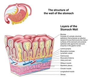
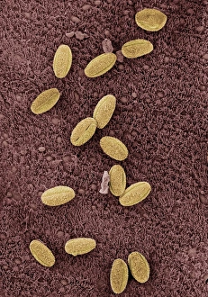
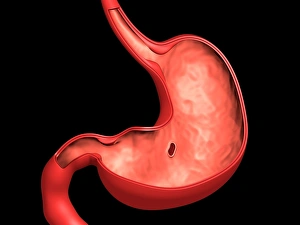
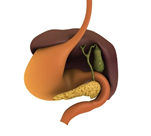
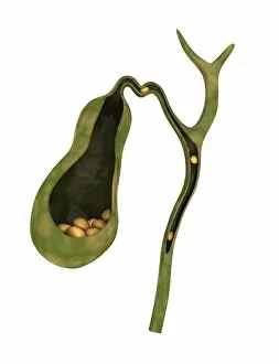
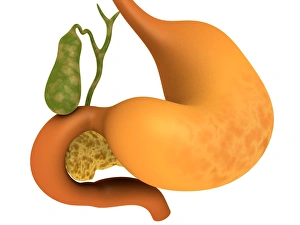
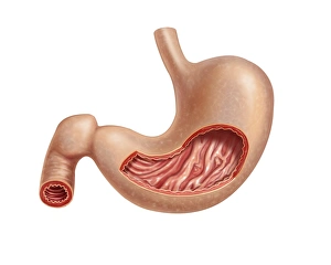
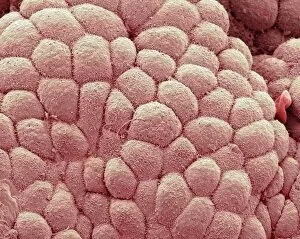
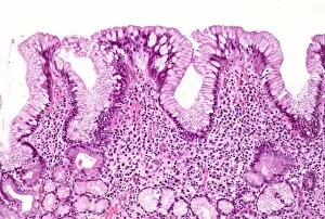
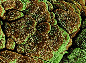
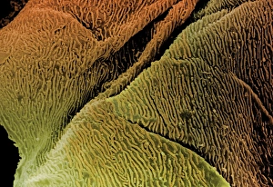
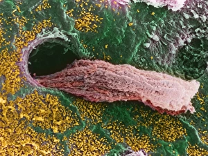
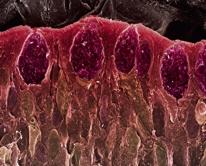
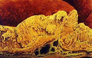
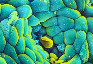
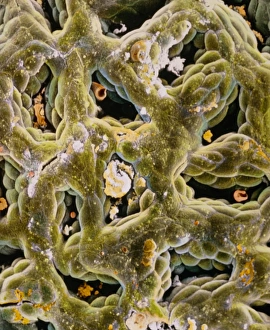
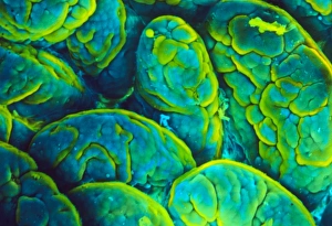
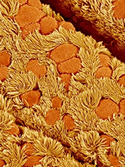
"Mucosa: The Versatile Lining of our Body's Vital Systems" The mucosa, a remarkable lining found in various parts of our body, plays an essential role in maintaining the health and functionality of several vital systems. From the nasal passages to the digestive tract, let's explore some intriguing aspects of this versatile tissue. Starting with the nasal lining, microscopic images captured through Scanning Electron Microscopy (SEM) reveal its intricate structure. This delicate mucosal layer not only filters out impurities but also warms and moisturizes the air we breathe. Moving on to the stomach, Gastric Antral Vascular Ectasia C016/8328 showcases a condition where blood vessels within the gastric mucosa become dilated. Understanding such anomalies helps medical professionals diagnose and treat related disorders effectively. Inhaling allergens can trigger allergic reactions within our trachea. SEM images provide a closer look at how these irritants interact with tracheal mucosa, leading to symptoms like coughing or wheezing. Cross-sections of both human stomach and large intestine illustrate their respective mucosal layers' importance in digestion and nutrient absorption. These illustrations help us comprehend how food interacts with these linings as it progresses through our digestive system. Conceptual images depicting peptic ulcers highlight one potential consequence when gastric mucosa becomes damaged due to factors like stress or bacterial infection. Such visuals emphasize why early detection and appropriate treatment are crucial for managing this condition effectively. When gallbladder-related issues arise, cholecystectomy may be necessary—the surgical removal of this organ responsible for storing bile produced by the liver. Illustrations showcasing this procedure shed light on how surgeons navigate around surrounding tissues while removing it safely. A conceptual image representing the entire human digestive system provides an overview of all organs involved—each equipped with its own specialized type of mucosal lining that contributes uniquely to overall digestion efficiency. Lastly, another conceptual image reveals the formation of gallstones within the gallbladder.