
Mucus Collection
"Mucus: A Sticky Substance with Vital Roles in the Human Body" Mucus, often associated with discomfort and illness


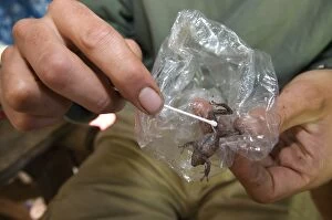


All Professionally Made to Order for Quick Shipping
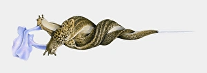
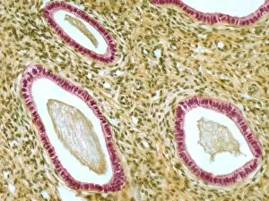
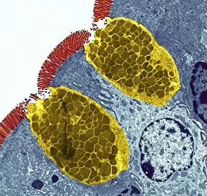
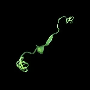
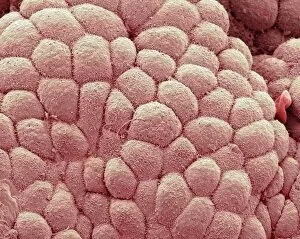
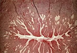
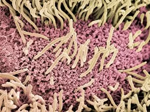
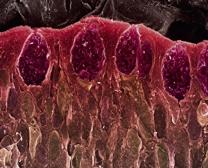
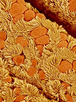
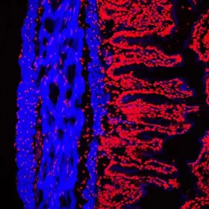
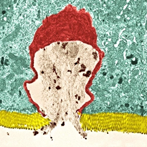
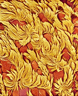
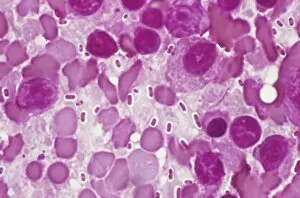
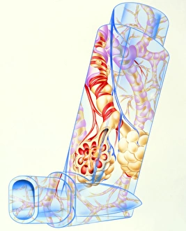
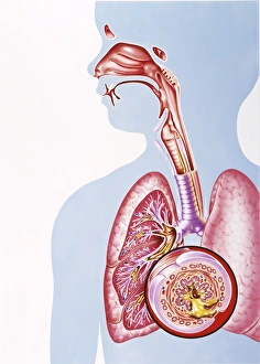
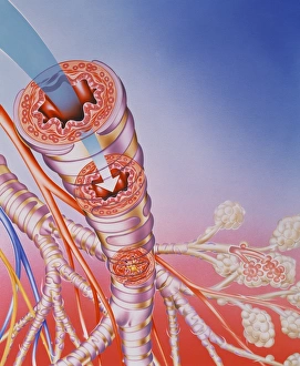
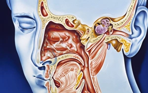
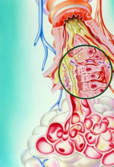
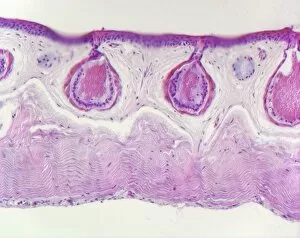
"Mucus: A Sticky Substance with Vital Roles in the Human Body" Mucus, often associated with discomfort and illness, plays a crucial role in various aspects of human physiology. From cystic fibrosis to the intricate workings of our reproductive system, mucus is an unsung hero that deserves recognition. In individuals with cystic fibrosis, a genetic disorder affecting the lungs and other organs, mucus becomes thick and sticky. This abnormal consistency hinders proper lung function, making breathing difficult and increasing susceptibility to infections. Moving away from the respiratory system, it also plays a significant role in female reproduction. The endometrium lining of the uterus is coated with a layer that changes throughout the menstrual cycle. This mucus helps facilitate sperm transport during ovulation while providing protection for fertilized eggs. The cervix acts as another gateway where mucus takes center stage. Light micrographs reveal its structure and composition, showcasing how it aids or restricts sperm movement depending on fertility status. Additionally, during pregnancy or impending labor, a protective barrier called the "mucus plug" forms within the cervix to prevent harmful bacteria from entering the uterus. As we delve deeper into our bodies' inner workings, we encounter more instances where mucus proves essential. In cross-sections of organs like the trachea or stomach lining, we witness how this slimy substance acts as a defense mechanism against foreign particles by trapping them before they can cause harm. Even during childbirth itself - an awe-inspiring process - mucus continues to play its part. Digital illustrations depict how as labor nears its peak stages; it's not only about contractions but also about cervical dilation facilitated by pressure from fetal head movements against both cervix and accompanying presence of that trusty "mucous plug. " Beyond reproductive matters lies yet another fascinating aspect involving nasal cavities and ciliate cells combating rhinovirus infections through their production and the presence of antibodies.