Fraud Blocker Skip to main content

Mycological Collection (page 3)

"Mycological Marvels

Background imageMycological Collection: Puffball (Lycoperdon perlatum) mushrooms C016 / 3434

Puffball (Lycoperdon perlatum) mushrooms C016 / 3434
Puffball (Lycoperdon perlatum) mushrooms, artwork

Background imageMycological Collection: Fairy ring (Marasmius oreades) mushrooms C016 / 3433

Fairy ring (Marasmius oreades) mushrooms C016 / 3433
Fairy ring (Marasmius oreades) mushrooms, artwork

Background imageMycological Collection: Lactarius deliciosus mushrooms, artwork C016 / 3367

Lactarius deliciosus mushrooms, artwork C016 / 3367
Saffron milk cap (Lactarius deliciosus) mushrooms, artwork

Background imageMycological Collection: Hygrophorus russula mushrooms, artwork C016 / 3364

Hygrophorus russula mushrooms, artwork C016 / 3364
Hygrophorus russula mushrooms, artwork

Background imageMycological Collection: Aspergillus fungus, artwork C013 / 4613

Aspergillus fungus, artwork C013 / 4613
Aspergillus fungus. Computer artwork of the fruiting body (conidiophore) of a typical mould (Aspergillus sp.), showing spores (conidia, round)

Background imageMycological Collection: Cells from a urine infection, SEM

Cells from a urine infection, SEM
Cells from a urine infection. Coloured scanning electron micrograph (SEM) of crenated red blood cells, white blood cells and epithelial cells present in a urine sample taken from someone with a

Background imageMycological Collection: Corn smut infection, light micrograph

Corn smut infection, light micrograph
Corn smut infection. Light micrograph of a section through tissue from a corn (Zea mays) plant infected with corn smut fungus (Ustilago maydis)

Background imageMycological Collection: Pear scab infection, light micrograph

Pear scab infection, light micrograph
Pear scab infection. Light micrograph of a section through a pear (Pyrus sp.) infected with the Venturia pirinum fungus. This fungus causes scab, or black spot, of the pear fruit

Background imageMycological Collection: Black scab infection, light micrograph

Black scab infection, light micrograph
Black scab infection. Light micrograph of a section through a potato infected with black scab (Synchytrium endobioticum) fungus

Background imageMycological Collection: Earth ball fungus, light micrograph

Earth ball fungus, light micrograph
Earth ball fungus. Light micrograph of a section through tissue from the fruiting body of an earth ball (Scleroderma vulgare) mushroom

Background imageMycological Collection: Plum pocket infection, light micrograph

Plum pocket infection, light micrograph
Plum pocket infection. Light micrograph of a section through a plum gall caused by the plum pocket fungus (Taphrina pruni)

Background imageMycological Collection: Boletus fungus, light micrograph

Boletus fungus, light micrograph
Boletus fungus. Light micrograph of a section through tissue from the fruiting body of a Boletus sp. mushroom, showing the hymenium (dark red) of the fruiting body

Background imageMycological Collection: Porcelain mushroom (Oudemansiella mucida)

Porcelain mushroom (Oudemansiella mucida)
Porcelain mushrooms (Oudemansiella mucida) growing on a moss-covered branch. This edible fungus is named after its translucent appearance. Photographed in the New Forest, UK, in October

Background imageMycological Collection: Neutrophil engulfing fungus, SEM

Neutrophil engulfing fungus, SEM
Neutrophil engulfing fungus. Coloured scanning electron micrograph (SEM) of a neutrophil white blood cell (orange) engulfing and destroying a Candida albicans (thrush) fungal hypha (green)

Background imageMycological Collection: Gill fungus (Lyophyllum decastes)

Gill fungus (Lyophyllum decastes) occurs in dense clusters. The word decastes means a company of ten men. The gills are covered in basidia bearing spores

Background imageMycological Collection: Beefsteak Fungus (Fistulina hepatica)

Beefsteak Fungus (Fistulina hepatica). This bracket fungus belonging to the homobasidiomycetes grows on the bark of living oak and sweet chestnut trees

Background imageMycological Collection: Magic mushrooms (Psilocybe semilanceata)

Magic mushrooms (Psilocybe semilanceata). These fungi, commonly known as liberty caps, contain the psychoactive compound psilocybin, which produces a hallucinogenic effect when ingested

Background imageMycological Collection: Tremella mesenterica

Tremella mesenterica
Yellow brain fungus (Tremella mesenterica) growing on a dead gorse bush. The fruiting body is soft, gelatinous and translucent with brain like lobes

Background imageMycological Collection: Common Earthball (Scleroderma citrinum)

Common Earthball (Scleroderma citrinum). The fruiting body is hemispherical with a tough outer layer covered in scales. Inside the ball is a purple-brown mass (gleba)

Background imageMycological Collection: Ladybird on fungus

Ladybird on fungus. Seven-spot ladybird (Coccinella septem-punctata) climbing up the stalk of a fungal fruiting body. This is a wood-rotting fungus growing on a dead tree stump

Background imageMycological Collection: Many-coloured polypore fungi

Many-coloured polypore fungi
Many-zoned polypore (Coriolus versicolor) bracket fungi. Wiltshire, UK November 2010

Background imageMycological Collection: Podoscypha fungus

Podoscypha fungus, New Forest, UK October 2009

Background imageMycological Collection: Parasol mushroom

Parasol mushroom (Macrolepiota procera). Photographed on Powerstock Common, Dorset, UK, in October

Background imageMycological Collection: Aspergillus fungus, SEM

Aspergillus fungus, SEM
Aspergillus fungus. Coloured scanning electron micrograph (SEM) of the fruiting body (round, top) of a typical mould (Aspergillus sp.)

Background imageMycological Collection: Parasol mushrooms

Parasol mushrooms (Macrolepiota procera). Photographed in Narke, Sweden

Background imageMycological Collection: Psathyrella mushrooms after forest fire

Psathyrella mushrooms after forest fire
Psathyrella pennata mushrooms growing one year after a forest fire. This fire took place in a clear-felled area in the Tiveden forest, in Vastra Gotaland County, Sweden

Background imageMycological Collection: Geopyxis elf-cup fungi after forest fire

Geopyxis elf-cup fungi after forest fire
Geopyxis carbonaria elf-cup fungi growing one year after a forest fire. This fire took place in a clear-felled area in the Tiveden forest, in Vastra Gotaland County, Sweden

Background imageMycological Collection: Honey fungus

Honey fungus (Armillaria mellea) growing on a tree trunk. Photogroahed in Narke, Sweden

Background imageMycological Collection: Jersey cow bolete

Jersey cow bolete (Suillus bovinus). Photographed in Narke, Sweden

Background imageMycological Collection: Myxomphalia mushroom after forest fire

Myxomphalia mushroom after forest fire
Myxomphalia maura mushrooms growing one year after a forest fire. This fire took place in a clear-felled area in the Tiveden forest, in Vastra Gotaland County, Sweden

Background imageMycological Collection: Helvella macropus mushroom

Helvella macropus mushroom
Helvella macropus fungus fruiting bodies. Photographed in Narke, Sweden

Background imageMycological Collection: Peziza cup fungus after forest fire

Peziza cup fungus after forest fire
Peziza cup fungus (Peziza lobulata) growing two months after a forest fire. This fire took place in a clear-felled area in the Tiveden forest, in Vastra Gotaland County, Sweden

Background imageMycological Collection: Plant regrowth after forest fire

Plant regrowth after forest fire
Plant regrowth one year after a forest fire. The plants and fungi seen here include Pholiota highlandensis mushrooms (lower left), the common liverwort (Marchantia polymorpha, white-green, centre)

Background imageMycological Collection: Birch bolete (Leccinum scabrum) mushroom

Birch bolete (Leccinum scabrum) mushroom
Brown birch bolete (Leccinum scabrum) mushroom growing amongst common heather (Calluna vulgaris). Photographed in the Cairngorms National Park, Scotland, in August

Background imageMycological Collection: Mushroom gill cap, light micrograph

Mushroom gill cap, light micrograph. Transverse section through the cap of a common mushroom (Psalliota sp.) showing the gill lamellae (white)

Background imageMycological Collection: Rust fungus infection, light micrograph

Rust fungus infection, light micrograph. Transverse section through a barberry (Berberis sp.) leaf infected by a parasitic rust (Puccinia sp.)

Background imageMycological Collection: Fly agaric (Amanita muscaria) mushrooms

Fly agaric (Amanita muscaria) mushrooms growing amongst common heather (Calluna vulgaris) in an ancient Caledonian Scots Pine woodland

Background imageMycological Collection: Mould fruiting bodies, light micrograph

Mould fruiting bodies, light micrograph. Black mould (Rhizopus nigricans) fruiting bodies (top and upper right) liberating spores

Background imageMycological Collection: Aspergillus fungus spores, SEM

Aspergillus fungus spores, SEM
Aspergillus fungus spores, coloured scanning electron micrograph (SEM). These chains of spores (orange) are the reproductive cells of the fruiting body, or conidiophore

Background imageMycological Collection: Amanita (Amanita frostiana) mushroom

Amanita (Amanita frostiana) mushroom
Frosts amanita (Amanita frostiana) mushroom. Photographed in Vermont, USA

Background imageMycological Collection: Woolly gomphus (Gomphus floccosus)

Woolly gomphus (Gomphus floccosus) mushroom. Photographed in Vermont, USA

Background imageMycological Collection: Edible fungi at a market

Edible fungi at a market. Selection of different mushrooms and fungal fruiting bodies arrayed for sale on a market stall. There are thousands of species of edible mushrooms found worldwide

Background imageMycological Collection: Puffball fungus

Puffball fungus (Lycoperdon foetidum)

Background imageMycological Collection: Penicillium fruiting bodies, SEM

Penicillium fruiting bodies, SEM

Background imageMycological Collection: Tar spot fungus on sycamore leaves

Tar spot fungus on sycamore leaves. Leaves of a sycamore (Acer pseudoplatanus) tree, affected by the fungal pathogen Rhytisma acerinum, which causes a disease known as tar spot

Background imageMycological Collection: Dried commercial yeast pellet, SEM

Dried commercial yeast pellet, SEM
Dried commercial yeast pellet. Coloured scanning electron micrograph (SEM) of a dried pellet of bakers yeast (Saccharomyces cerevisiae). Yeast is a fungus and consists of single vegetative cells. S

Background imageMycological Collection: Map lichen (Rhizocarpon geographicum)

Map lichen (Rhizocarpon geographicum) on a rock. Lichens are symbiotic organisms formed of a fungus and an alga that mutually benefit one another. Photographed in Scotland

Background imageMycological Collection: Lichen (Cladonia stellaris)

Lichen (Cladonia stellaris)
Cladonia stellaris lichen (white) and lingonberry (Vaccinium vitis-idaea, green and orange). Lichens are symbiotic organisms formed of a fungus and an alga that mutually benefit one another


All Professionally Made to Order for Quick Shipping

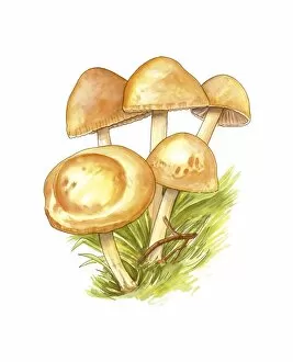
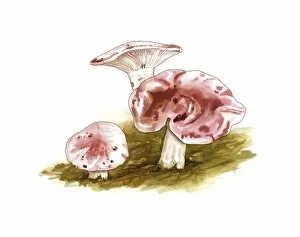
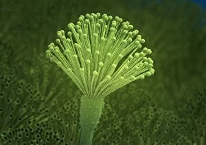
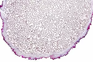
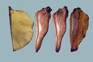
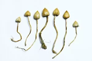
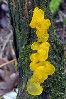
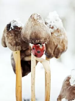
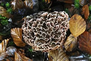
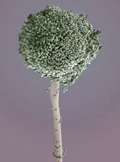
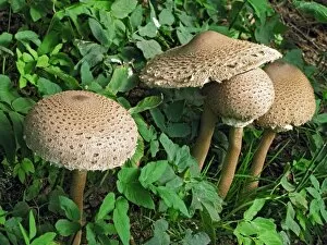
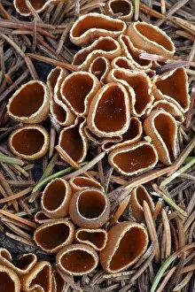
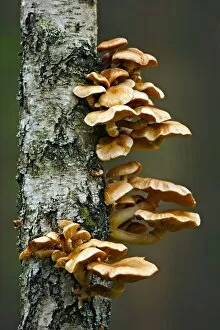
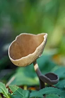
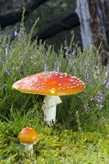
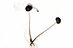
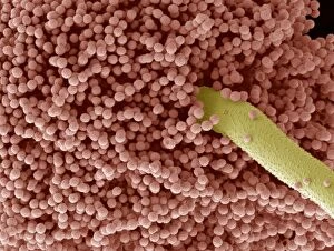
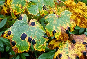
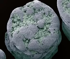
"Mycological Marvels: Exploring the Fascinating World of Fungi" Step into a realm where bread mould becomes a captivating subject under the scanning electron microscope (SEM). Witness the intricate details and mesmerizing patterns that adorn these seemingly ordinary fungi. Transport yourself to the early 1900s, as you gaze upon a botanical plate depicting both Good and Bad Mushrooms. Delight in the vibrant colors captured through lithography, showcasing nature's diversity in mushroom species. Discover Hyprophyllum aquifolii, elegantly displayed on Plate 38 from Iconographie des Champignons de J. J. , its delicate form beckoning you closer. Admire Hypodendrums fagi and queris on Plate 138, their unique features highlighted with precision by J. J. 's artistic hand. Marvel at Hypophyllum campestre or the field mushroom on Plate 130 from 'Iconographie des Champignons de J. J. , ' an exquisite representation of this culinary delight found in meadows. Let your imagination run wild as you envision Tubiporus cepa on Plate 176, enticingly illustrated for Traite des Champignons', promising a feast for those who dare to indulge. Delve deeper into Tubiporus esculentus on Plate 168 from Iconographie des Champignons de J. J. , marveling at its edible allure and contemplating its potential uses in gastronomy. Witness fungal fruiting bodies come alive before your eyes through stunning illustrations that capture their beauty and complexity. Explore another Botanical plate depicting Good and Bad Mushrooms from c. 1900 - a testament to mankind's fascination with these enigmatic organisms throughout history. Uncover Rust fungus infection through SEM imagery, revealing both the destructive power and awe-inspiring intricacy of nature's battles within microscopic realms. And finally, ponder upon a mouldy lemon.