Fraud Blocker Skip to main content

Mycological Collection (page 2)

"Mycological Marvels

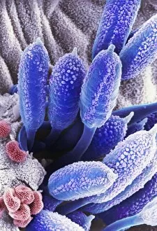
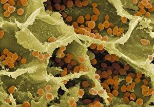
Background imageMycological Collection: Rust fungus on a rose leaf, SEM C017 / 7130

Rust fungus on a rose leaf, SEM C017 / 7130
Rust fungus on a rose leaf. Coloured scanning electron micrograph (SEM) of rust fungus (Phragmidium sp.) spores emerging from a rose (Rosa sp.) leaf (mauve)

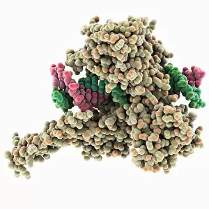
Background imageMycological Collection: Type I topoisomerase bound to DNA C014 / 0862

Type I topoisomerase bound to DNA C014 / 0862
Type I topoisomerase bound to DNA. Molecular model showing a type I topoisomerase molecule (khaki) bound to a strand of DNA (deoxyribonucleic acid, pink and green)

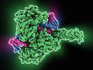
Background imageMycological Collection: Type I topoisomerase bound to DNA C014 / 0883

Type I topoisomerase bound to DNA C014 / 0883
Type I topoisomerase bound to DNA. Molecular model showing a type I topoisomerase molecule (green) bound to a strand of DNA (deoxyribonucleic acid, pink and blue)

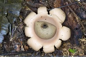
Background imageMycological Collection: Earthstar (Geastrum sp. ) fungus C014 / 0950

Earthstar (Geastrum sp. ) fungus C014 / 0950
Earthstar (Geastrum sp.) fungus in leaf litter. This fungus releases its spores from the central spherical structure. Photographed in Rio Bigal Biological Reserve, Amazon Rainforest, Ecuador

Background imageMycological Collection: Parasitic fungus on a weevil C014 / 0951

Parasitic fungus on a weevil C014 / 0951
Parasitic fungus on a weevil. Fruiting body of a Cordyceps sp. fungus (left) growing out of the body of a weevil (superfamily Curculionoidea, right) clinging to a plant stem

Background imageMycological Collection: Mushrooms, historical model

Mushrooms, historical model. 1880 papier mache models of different types of mushrooms

Background imageMycological Collection: Penicillium colony C014 / 4861

Penicillium colony C014 / 4861
Penicillium colony. Colony of Penicillium chrysogenum fungus growing on agar in a petri dish. This fungus produces the antibiotic penicillin G, seen as small droplets on the surface

Background imageMycological Collection: Penicillium colony C014 / 4857

Penicillium colony C014 / 4857
Penicillium colony. Colony of Penicillium chrysogenum fungus growing on agar in a petri dish. This fungus produces the antibiotic penicillin G, seen as small droplets on the surface

Background imageMycological Collection: Lichen, SEM C014 / 4736

Lichen, SEM C014 / 4736
Lichen. Coloured scanning electron micrograph (SEM) showing the structure of a lichen. Lichens are the result of a symbiotic relationship between a fungus and an alga

Background imageMycological Collection: Yeast on a grape, SEM C014 / 4746

Yeast on a grape, SEM C014 / 4746
Yeast on a grape. Coloured scanning electron micrograph (SEM) of yeast cells (red) on the skin of a grape. Magnification: x1250 when printed 10 cm wide

Background imageMycological Collection: Fungal spores, SEM C014 / 4742

Fungal spores, SEM C014 / 4742
Fungal spores. Coloured scanning electron micrograph (SEM) of a section through a sporangium (green) from a Tranzschelia fusca fungus, showing the spores (pink) inside

Background imageMycological Collection: Dotted stem bolete (Boletus erythropus) C014 / 0740

Dotted stem bolete (Boletus erythropus) C014 / 0740
Dotted stem bolete (Boletus erythropus) mushroom cut in half to show its interior. This mushroom starts off yellow inside

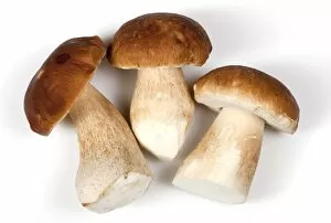
Background imageMycological Collection: Porcini (Boletus edulis) mushroom C014 / 0738

Porcini (Boletus edulis) mushroom C014 / 0738
Porcini (Boletus edulis) mushroom

Background imageMycological Collection: Porcini (Boletus edulis) mushrooms C014 / 0737

Porcini (Boletus edulis) mushrooms C014 / 0737
Porcini (Boletus edulis) mushrooms

Background imageMycological Collection: Porcini (Boletus edulis) mushrooms C014 / 0736

Porcini (Boletus edulis) mushrooms C014 / 0736
Porcini (Boletus edulis) mushrooms

Background imageMycological Collection: Pine bolete (Boletus pinophilus) C014 / 0735

Pine bolete (Boletus pinophilus) C014 / 0735
Pine bolete (Boletus pinophilus) mushroom, cut in half

Background imageMycological Collection: Coral fungus on rainforest floor C016 / 8425

Coral fungus on rainforest floor C016 / 8425
Coral fungus on rainforest floor. Strict-branch coral (Ramaria stricta) fungus growing on dead wood in the floor of a rainforest. Photographed in Napo Province, Amazon rainforest, Ecuador

Background imageMycological Collection: Diseased cocoa pods, Ecuador C016 / 8421

Diseased cocoa pods, Ecuador C016 / 8421
Diseased cocoa pods. Cocoa (Theobroma cacao) fruit (pods) infected with frosty pod rot disease. This damaging disease is caused by the basidiomycete fungus Moniliophthora roreri

Background imageMycological Collection: Pine bolete (Boletus pinophilus) C014 / 0734

Pine bolete (Boletus pinophilus) C014 / 0734
Pine bolete (Boletus pinophilus) mushroom

Background imageMycological Collection: Cicada infected with fungus C014 / 4581

Cicada infected with fungus C014 / 4581
Cicada infected with fungus. Close-up of a dead cicada (family Cicadidae) that has been killed by a parasitic fungus

Background imageMycological Collection: Cicada infected with fungus C014 / 4582

Cicada infected with fungus C014 / 4582
Cicada infected with fungus. Dead cicada (family Cicadidae) that has been killed by a parasitic fungus

Background imageMycological Collection: Cicada infected with fungus C014 / 4580

Cicada infected with fungus C014 / 4580
Cicada infected with fungus. Dead cicada (family Cicadidae) that has been killed by a parasitic fungus

Background imageMycological Collection: Morel (Mitrophora semilibera) mushrooms C014 / 0699

Morel (Mitrophora semilibera) mushrooms C014 / 0699
Half-free morel (Mitrophora semilibera) mushrooms

Background imageMycological Collection: Morel (Mitrophora semilibera) mushroom C014 / 0697

Morel (Mitrophora semilibera) mushroom C014 / 0697
Half-free morel (Mitrophora semilibera) mushroom

Background imageMycological Collection: Fungus parasitising a fly

Fungus parasitising a fly. Close-up of a fly that has been killed by the parasitic fungus Entomophthora muscae. This fungus is highly adapted to its host

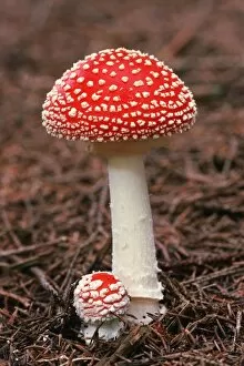
Background imageMycological Collection: Fly agaric (Amanita muscaria) fungus C014 / 3019

Fly agaric (Amanita muscaria) fungus C014 / 3019
Fly agaric (Amanita muscaria) fungus. These poisonous mushrooms are the fruiting bodies of the Amanita muscaria fungus. The caps have long been dried

Background imageMycological Collection: Fungal fruiting bodies, 19th century C015 / 6087

Fungal fruiting bodies, 19th century C015 / 6087
Fungal fruiting bodies, 19th-century artwork. The shape of fungal fruiting bodies can range from capped and gilled toadstools and mushrooms (top), to bracket fungi (upper centre), morels (centre)

Background imageMycological Collection: Mycena epipterygia mushroom C018 / 2535

Mycena epipterygia mushroom C018 / 2535
Mycena epipterygia mushroom

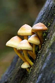
Background imageMycological Collection: Sulphur tuft (Hypholoma fasciculare) C018 / 2565

Sulphur tuft (Hypholoma fasciculare) C018 / 2565
Sulphur tuft (Hypholoma fasciculare) mushrooms

Background imageMycological Collection: Sulphur tuft (Hypholoma fasciculare) C018 / 2534

Sulphur tuft (Hypholoma fasciculare) C018 / 2534
Sulphur tuft (Hypholoma fasciculare) mushrooms

Background imageMycological Collection: Scleroderma citrinum mushroom C018 / 2538

Scleroderma citrinum mushroom C018 / 2538
Common earthball (Scleroderma citrinum) mushroom

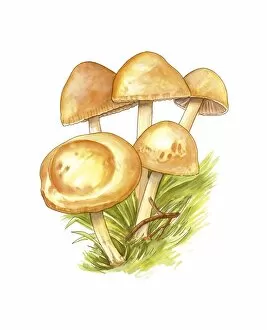
Background imageMycological Collection: Shaggy pholiota (Pholiota aurivella) C018 / 2566

Shaggy pholiota (Pholiota aurivella) C018 / 2566
Shaggy pholiota (Pholiota aurivella) mushrooms

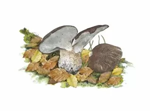
Background imageMycological Collection: Beechwood sickener (Russula nobilis) C018 / 2532

Beechwood sickener (Russula nobilis) C018 / 2532
Beechwood sickener (Russula nobilis) mushroom

Background imageMycological Collection: Shaggy pholiota (Pholiota aurivella) C018 / 2533

Shaggy pholiota (Pholiota aurivella) C018 / 2533
Shaggy pholiota (Pholiota aurivella) mushrooms

Background imageMycological Collection: Fungus on a redwood tree C018 / 2574

Fungus on a redwood tree C018 / 2574
Fungus on a redwood tree. Tree fungus growing on a coast redwood (Sequoia sempervirens) tree. Photographed in Muir Woods National Park, California, USA

Background imageMycological Collection: Hypholoma mushrooms C018 / 2568

Hypholoma mushrooms C018 / 2568
Hypholoma mushrooms. Sulphur tuft (Hypholoma fasciculare, left) and brick cap (Hypholoma sublateritium, right) mushrooms growing on a tree stump

Background imageMycological Collection: Porcelain fungus (Oudemansiella mucida) C018 / 2564

Porcelain fungus (Oudemansiella mucida) C018 / 2564
Porcelain fungus (Oudemansiella mucida) mushroom

Background imageMycological Collection: Mycena epipterygia mushroom C018 / 2536

Mycena epipterygia mushroom C018 / 2536
Mycena epipterygia mushroom

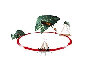
Background imageMycological Collection: Zombie ant fungus life-cycle, artwork

Zombie ant fungus life-cycle, artwork
Zombie ant fungus life-cycle. Computer artwork showing the life cycle of the parasitoidal fungus Ophiocordyceps unilateralis

Background imageMycological Collection: Scutiger pes-caprae-sabatera mushrooms C016 / 3446

Scutiger pes-caprae-sabatera mushrooms C016 / 3446
Scutiger pes-caprae-sabatera mushrooms, artwork

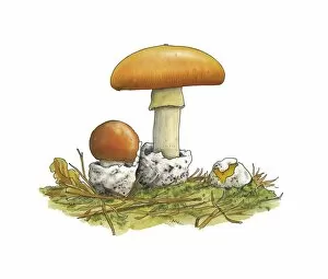
Background imageMycological Collection: Caesars mushrooms (Amanita caesarea)

Caesars mushrooms (Amanita caesarea), artwork

Background imageMycological Collection: Puffball (Lycoperdon perlatum) mushrooms C016 / 3434

Puffball (Lycoperdon perlatum) mushrooms C016 / 3434
Puffball (Lycoperdon perlatum) mushrooms, artwork

Background imageMycological Collection: Fairy ring (Marasmius oreades) mushrooms C016 / 3433

Fairy ring (Marasmius oreades) mushrooms C016 / 3433
Fairy ring (Marasmius oreades) mushrooms, artwork

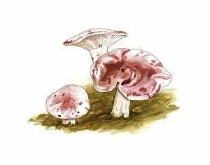
Background imageMycological Collection: Lactarius deliciosus mushrooms, artwork C016 / 3367

Lactarius deliciosus mushrooms, artwork C016 / 3367
Saffron milk cap (Lactarius deliciosus) mushrooms, artwork

Background imageMycological Collection: Hygrophorus russula mushrooms, artwork C016 / 3364

Hygrophorus russula mushrooms, artwork C016 / 3364
Hygrophorus russula mushrooms, artwork

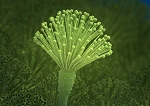
Background imageMycological Collection: Aspergillus fungus, artwork C013 / 4613

Aspergillus fungus, artwork C013 / 4613
Aspergillus fungus. Computer artwork of the fruiting body (conidiophore) of a typical mould (Aspergillus sp.), showing spores (conidia, round)

Background imageMycological Collection: Cells from a urine infection, SEM

Cells from a urine infection, SEM
Cells from a urine infection. Coloured scanning electron micrograph (SEM) of crenated red blood cells, white blood cells and epithelial cells present in a urine sample taken from someone with a

Background imageMycological Collection: Corn smut infection, light micrograph

Corn smut infection, light micrograph
Corn smut infection. Light micrograph of a section through tissue from a corn (Zea mays) plant infected with corn smut fungus (Ustilago maydis)


All Professionally Made to Order for Quick Shipping

"Mycological Marvels: Exploring the Fascinating World of Fungi" Step into a realm where bread mould becomes a captivating subject under the scanning electron microscope (SEM). Witness the intricate details and mesmerizing patterns that adorn these seemingly ordinary fungi. Transport yourself to the early 1900s, as you gaze upon a botanical plate depicting both Good and Bad Mushrooms. Delight in the vibrant colors captured through lithography, showcasing nature's diversity in mushroom species. Discover Hyprophyllum aquifolii, elegantly displayed on Plate 38 from Iconographie des Champignons de J. J. , its delicate form beckoning you closer. Admire Hypodendrums fagi and queris on Plate 138, their unique features highlighted with precision by J. J. 's artistic hand. Marvel at Hypophyllum campestre or the field mushroom on Plate 130 from 'Iconographie des Champignons de J. J. , ' an exquisite representation of this culinary delight found in meadows. Let your imagination run wild as you envision Tubiporus cepa on Plate 176, enticingly illustrated for Traite des Champignons', promising a feast for those who dare to indulge. Delve deeper into Tubiporus esculentus on Plate 168 from Iconographie des Champignons de J. J. , marveling at its edible allure and contemplating its potential uses in gastronomy. Witness fungal fruiting bodies come alive before your eyes through stunning illustrations that capture their beauty and complexity. Explore another Botanical plate depicting Good and Bad Mushrooms from c. 1900 - a testament to mankind's fascination with these enigmatic organisms throughout history. Uncover Rust fungus infection through SEM imagery, revealing both the destructive power and awe-inspiring intricacy of nature's battles within microscopic realms. And finally, ponder upon a mouldy lemon.