Home > Science > SEM
Rust fungus on a rose leaf, SEM C017 / 7130

![]()
Wall Art and Photo Gifts from Science Photo Library
Rust fungus on a rose leaf, SEM C017 / 7130
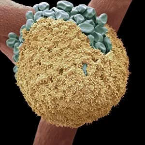
Rust fungus on a rose leaf. Coloured scanning electron micrograph (SEM) of rust fungus (Phragmidium sp.) spores emerging from a rose (Rosa sp.) leaf (mauve). Spores are the reproductive cells of a fungus. Two types of spore are seen, the over-wintering teleutospores (blue) and the summer uredospores (red). Rust fungi are parasitic on both wild and cultivated plants, often causing severe disease. Magnification: x440 when printed at 10 centimetres tall
Science Photo Library features Science and Medical images including photos and illustrations
Media ID 9232841
© MICROFIELD SCIENTIFIC LTD/SCIENCE PHOTO LIBRARY
Emerging Fungal Fungi Fungus Microbiology Mycological Mycology Plant Disease Plant Pathology Reproduction Reproductive Cell Rosa Rose Rust Spores Microbiological Pathogen
EDITORS COMMENTS
This print showcases the intricate world of rust fungus on a delicate rose leaf. In this coloured scanning electron micrograph (SEM), we witness the emergence of spores from the leaf's surface, revealing the reproductive cells of this parasitic Phragmidium sp. fungus. The contrasting hues highlight two distinct types of spores: teleutospores, responsible for over-wintering and depicted in striking blue, and uredospores, vibrant in red as they emerge during summer. Rust fungi are notorious pathogens that afflict both wild and cultivated plants, often causing severe diseases. This image offers a glimpse into their biological mechanisms at play. With a magnification of x440 when printed at 10 centimetres tall, it allows us to appreciate the microscopic intricacies involved in fungal reproduction. The mesmerizing beauty captured here serves as a reminder that even within nature's smallest realms lies an astonishing complexity waiting to be explored. As we delve into mycology and plant pathology through this photograph, we gain insight into how these tiny organisms impact our environment. Microfield Scientific Ltd/Science Photo Library presents us with yet another stunning visual representation bridging science and art—a testament to their dedication in capturing scientific wonders for all to behold.
MADE IN AUSTRALIA
Safe Shipping with 30 Day Money Back Guarantee
FREE PERSONALISATION*
We are proud to offer a range of customisation features including Personalised Captions, Color Filters and Picture Zoom Tools
SECURE PAYMENTS
We happily accept a wide range of payment options so you can pay for the things you need in the way that is most convenient for you
* Options may vary by product and licensing agreement. Zoomed Pictures can be adjusted in the Cart.