
Necrosis Collection
"Necrosis: Unveiling the Dark Side of Skin Disorders and Artwork" Step into the intriguing world of necrosis


All Professionally Made to Order for Quick Shipping
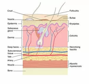
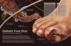
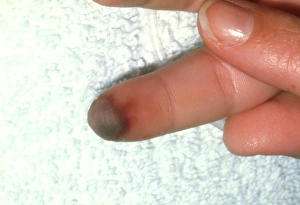
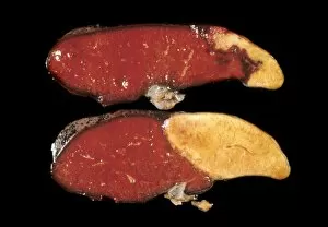
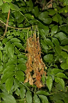
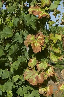
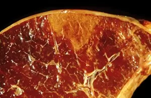
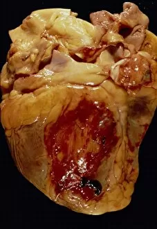
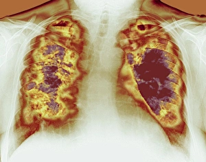
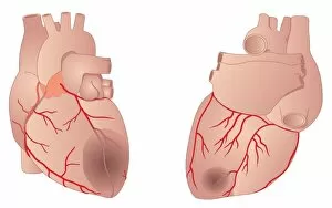
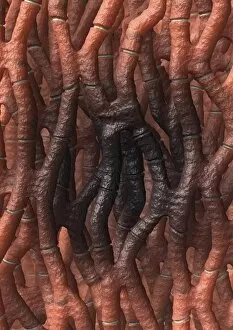
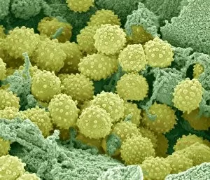
"Necrosis: Unveiling the Dark Side of Skin Disorders and Artwork" Step into the intriguing world of necrosis, where skin disorders and artwork intertwine to create a hauntingly beautiful narrative. In this captivating journey, we explore various manifestations across different mediums. One such masterpiece is "Profile of a Woman with Necrosis of the Nose" by Louis-Auguste Bisson. Painted between 1841-48, it depicts the devastating impact of necrotic tissue on facial features, evoking both fascination and empathy. Histopathology unravels the pathophysiology behind diabetic foot ulcers, showcasing how compromised blood flow leads to peripheral vasoconstriction in patients' fingers. The image captures not only physical deterioration but also serves as a poignant reminder of the silent struggles faced by those affected. Secondary period syphilis symptoms leave an indelible mark on bodies, revealing disfiguring lesions that result from untreated infections. This visual representation reminds us of the importance of early diagnosis and treatment in preventing further damage. Nature too falls victim to necrotic diseases like Ash Dieback caused by Chalara fraxinea fungal disease. Common Ash trees bear witness to their own demise as leaves wither away due to this relentless affliction in Norfolk. Splenic infarct C015/6220 showcases internal organ decay resulting from compromised blood supply. It highlights how even our vital organs are susceptible to necrotic events that can have severe consequences for overall health. Microscopic exploration reveals lymphocytes within hair follicles undergoing necrosis under scanning electron microscopy (SEM). This intricate view offers insight into cellular processes gone awry and emphasizes the delicate balance required for healthy functioning. The removal of an entire elbow joint due to humerus necrosis sheds light on extreme cases where surgical intervention becomes necessary for survival or improved quality of life. A stark reminder that sometimes drastic measures must be taken when confronted with necrotic conditions.