Fraud Blocker Skip to main content

Nerve Cells Collection

Nerve cells, also known as neurons, are the building blocks of our intricate nervous system

Background imageNerve Cells Collection: Motor neurons, light micrograph

Motor neurons, light micrograph. Motor neurons are responsible for passing information around the central nervous system (CNS) and from the CNS to the rest of the body

Background imageNerve Cells Collection: The microscopic structure of cells

The microscopic structure of cells
2626843 The microscopic structure of cells; (add.info.: Rudolph Virchow (1821-1902) is regarded as perhaps the greatest medical scientist of the nineteenth century

Background imageNerve Cells Collection: Santiago Ramon y Cajal, histologist

Santiago Ramon y Cajal, histologist
Santiago Ramon y Cajal (1852-1934), Spanish histologist. Ramon y Cajal was apprenticed to a barber and a shoemaker before taking up medicine

Background imageNerve Cells Collection: Anatomical pathways of innervation to the lacrimal gland

Anatomical pathways of innervation to the lacrimal gland

Background imageNerve Cells Collection: Conceptual image of synapse of neuron inside brain

Conceptual image of synapse of neuron inside brain

Background imageNerve Cells Collection: Microscopic view of multiple nerve cells, known as neurons

Microscopic view of multiple nerve cells, known as neurons
Microscopic view of multiple nerve cells, which are also called neurons. These are responsible for passing information around the central nervous system within the human body

Background imageNerve Cells Collection: Microscopic view of a unipolar neuron

Microscopic view of a unipolar neuron. A unipolar neuron is a type of neuron in which only one protoplasmic process (neurite) extends from the cell body

Background imageNerve Cells Collection: Detail of a nerve bundle

Detail of a nerve bundle

Background imageNerve Cells Collection: Orbital cut showing abducent nerve with ciliary ganglion and oculomotor nerve

Orbital cut showing abducent nerve with ciliary ganglion and oculomotor nerve

Background imageNerve Cells Collection: Representation of how our senses affect our thoughts

Representation of how our senses affect our thoughts

Background imageNerve Cells Collection: Conceptual image of synaptic vesicles

Conceptual image of synaptic vesicles

Background imageNerve Cells Collection: Conceptual image of human brain with neurons

Conceptual image of human brain with neurons in background

Background imageNerve Cells Collection: Nerve ending, seen in lower right, sends pain message from injured muscle

Nerve ending, seen in lower right, sends pain message from injured muscle. Blood vessel and immune cells are seen in the center and upper right of image

Background imageNerve Cells Collection: Representation of how our thoughts affect our emotions

Representation of how our thoughts affect our emotions

Background imageNerve Cells Collection: A nerve synapse showing the release of neurotransmitters

A nerve synapse showing the release of neurotransmitters
Detail of a nerve synapse showing the release of neurotransmitters

Background imageNerve Cells Collection: Microscopic view of nerve fibers

Microscopic view of nerve fibers. A nerve fiber is a threadlike extension of a nerve cell in the nervous system

Background imageNerve Cells Collection: Anatomy structure of neurons

Anatomy structure of neurons

Background imageNerve Cells Collection: Conceptual image of synapse receptors

Conceptual image of synapse receptors

Background imageNerve Cells Collection: Schematic of the hypothalamus receiving nerve impulses from the body

Schematic of the hypothalamus receiving nerve impulses from the body and sending messages to the circulatory and nervous system

Background imageNerve Cells Collection: Human brain showing the layout of the sensory cortex

Human brain showing the layout of the sensory cortex
Coronal section through human brain showing the layout of the sensory cortex

Background imageNerve Cells Collection: Conceptual image of synaptic transmission

Conceptual image of synaptic transmission

Background imageNerve Cells Collection: Microscopic view of a bipolar neuron

Microscopic view of a bipolar neuron. A bipolar cell is a type of neuron which has two extensions. Bipolar cells are specialized sensory neurons for the transmission of special senses

Background imageNerve Cells Collection: Close-up view of the synapse in the nervous system

Close-up view of the synapse in the nervous system. A synapse is a structure that permits a neuron (or nerve cell) to pass an electrical or chemical signal to another cell

Background imageNerve Cells Collection: Primary cultures of superior cervical ganglia cells

Primary cultures of superior cervical ganglia cells

Background imageNerve Cells Collection: Nerve with myelin sheath, seen in lower right, connects with muscle

Nerve with myelin sheath, seen in lower right, connects with muscle. Blood vessel and immune cells are seen in the center and upper right of image

Background imageNerve Cells Collection: Abducent nerve in the orbit with lateral rectus muscle

Abducent nerve in the orbit with lateral rectus muscle

Background imageNerve Cells Collection: Conceptual image of GABA receptors

Conceptual image of GABA receptors. The GABA receptors are a class of receptors that respond to the neurotransmitter gamma-aminobutyric acid

Background imageNerve Cells Collection: Conceptual image of a neuron ligntning signal passing

Conceptual image of a neuron ligntning signal passing

Background imageNerve Cells Collection: Microscopic view of inner nervous system

Microscopic view of inner nervous system

Background imageNerve Cells Collection: Microscopic view of a multipolar neuron

Microscopic view of a multipolar neuron. Multipolar neurons possess a single axon and many dendrites

Background imageNerve Cells Collection: Illustration of action potential of a nerve cell

Illustration of action potential of a nerve cell

Background imageNerve Cells Collection: Conceptual image of a neuron

Conceptual image of a neuron

Background imageNerve Cells Collection: Pyramidal nerve cells, artwork C017 / 2273

Pyramidal nerve cells, artwork C017 / 2273
Pyramidal nerve cells. Computer artwork of pyramidal nerve cells from the cerebral cortex of the brain. Pyramidal cells are so named for their triangular cell bodies

Background imageNerve Cells Collection: Pyramidal nerve cells, artwork C017 / 2274

Pyramidal nerve cells, artwork C017 / 2274
Pyramidal nerve cells. Computer artwork of pyramidal nerve cells from the cerebral cortex of the brain. Pyramidal cells are so named for their triangular cell bodies

Background imageNerve Cells Collection: Myelin sheats and glial cells, artwork C014 / 2646

Myelin sheats and glial cells, artwork C014 / 2646
Computer artwork depicting axons surrounded by a myelin sheath (brown) and microglial cells (light blue). Glial cells are nervous system cells that provide structural support

Background imageNerve Cells Collection: Myelin sheats and glial cells, artwork C014 / 2647

Myelin sheats and glial cells, artwork C014 / 2647
Computer artwork depicting axons surrounded by a myelin sheath (brown) and microglial cells (light blue). Glial cells are nervous system cells that provide structural support

Background imageNerve Cells Collection: Cerebrum, light micrograph C014 / 0636

Cerebrum, light micrograph C014 / 0636
Cerebrum. Light micrograph of a section through part of the cerebrum of the brain. Seen here are the three main layers. The outer grey matter comprises the molecular layer (light pink)

Background imageNerve Cells Collection: Retina blood vessel and nerve cells

Retina blood vessel and nerve cells
Retina cells. Fluorescent light micrograph of cells in the retina, the light-sensitive membrane that lines the back of the eyeball

Background imageNerve Cells Collection: Immunofluorescent LM of rat brain cells and axons

Immunofluorescent LM of rat brain cells and axons
Cortical brain cells and axons. Immunofluorescent Light Micrograph of rat cortical brain cells and axons growing in culture

Background imageNerve Cells Collection: Spinal nerve ganglion, light micrograph

Spinal nerve ganglion, light micrograph
Spinal nerve ganglion. Light micrograph of a cross-section through a spinal nerve ganglion. This is a node of nerve cells located just outside the spinal cord at the point where it is joined by

Background imageNerve Cells Collection: Synapse, computer artwork

Synapse, computer artwork
Synapses. Computer artwork of synapses, the junctions between the ends (blue, swollen) of two nerve cells (neurons). Nerve cells are responsible for passing information around the central nervous

Background imageNerve Cells Collection: Nerve cell growth

Nerve cell growth. Light micrograph of nerve cells (neurons) with immunofluorescent staining. These cells have been grown in culture

Background imageNerve Cells Collection: Cell death

Cell death. Computer-enhanced confocal light micrograph of cells in the retina of the eye undergoing programmed cell death (apoptosis)

Background imageNerve Cells Collection: Alzheimers disease

Alzheimers disease
Conceptual computer artwork depicting Alzheimers disease and other brain disease

Background imageNerve Cells Collection: Pyramidal neurons, light micrograph

Pyramidal neurons, light micrograph with Nissl staining. This sample, from a human brain, shows two pyramidal neurons. Pyramidal neurons (nerve cells)

Background imageNerve Cells Collection: Purkinje neurons, light micrograph

Purkinje neurons, light micrograph

Background imageNerve Cells Collection: Neural network

Neural network. Artwork of nerve cells (neurons, green) connected by nerve processes (dendrites and axons) to form a neural network

Background imageNerve Cells Collection: Neuromuscular junctions, artwork

Neuromuscular junctions, artwork
Neuromuscular junctions. Computer artwork of junctions between nerves (thread-like objects) and a muscle (purple surface)


All Professionally Made to Order for Quick Shipping

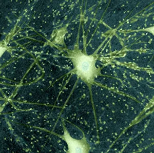
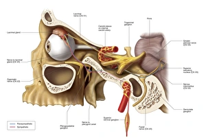
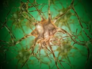
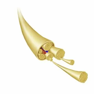
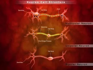
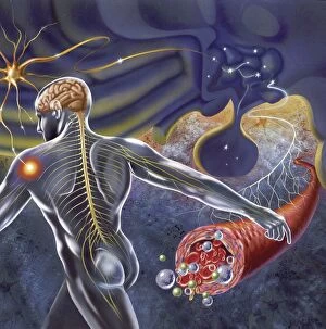
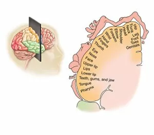
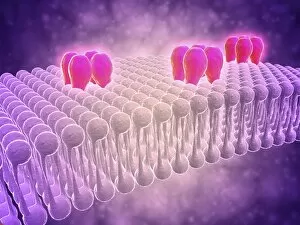
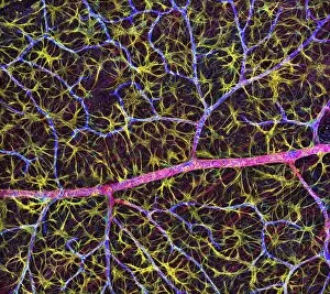
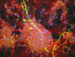
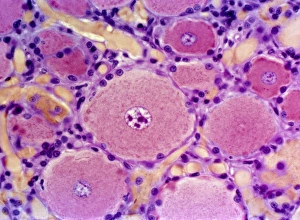
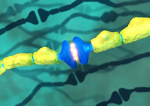
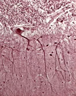
Nerve cells, also known as neurons, are the building blocks of our intricate nervous system. These remarkable cells play a crucial role in transmitting signals throughout our body, allowing us to move, think, and feel. Motor neurons are a type of nerve cell that carry messages from the brain to muscles or glands. Through their long extensions called axons, they enable precise movements and coordinated actions. Santiago Ramon y Cajal, a renowned histologist, made significant contributions to our understanding of nerve cells. His detailed light micrographs revealed the complex structure and organization of these cells within the brain. Anatomical pathways of innervation to the lacrimal gland demonstrate how nerve fibers connect with this tear-producing gland. This connection ensures proper lubrication for our eyes. Primary cultures of superior cervical ganglia cells provide researchers with valuable insights into how these specific groups function and communicate with each other. A conceptual image depicting synapses inside the brain showcases the incredible complexity involved in transmitting information between neurons. These tiny gaps allow electrical impulses to jump from one neuron to another through chemical messengers called neurotransmitters. Microscopic views offer glimpses into multiple nerve cells or neurons at work. They reveal their unique shapes and structures as they form networks responsible for relaying information throughout our body. One such view highlights a unipolar neuron - a type characterized by having only one extension emerging from its cell body. This specialized structure allows efficient transmission over long distances within our nervous system. The detail captured in an image showcasing a nerve bundle emphasizes their importance in carrying signals across various regions of our body rapidly. Bundles like these ensure effective communication between different parts of our anatomy. Another microscopic view captures multiple interconnected neurons once again – emphasizing their abundance and essential role in maintaining overall bodily functions seamlessly. An orbital cut displaying nerves like abducent nerves alongside ciliary ganglion and oculomotor nerves reveals how intricately woven these neural pathways are. They enable precise eye movements and coordination.