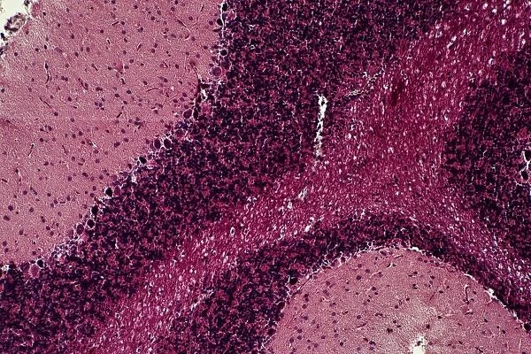
Cerebrum, light micrograph C014 / 0636
![]()

Wall Art and Photo Gifts from Science Photo Library
Cerebrum, light micrograph C014 / 0636
Cerebrum. Light micrograph of a section through part of the cerebrum of the brain. Seen here are the three main layers. The outer grey matter comprises the molecular layer (light pink) and the granular layer (purple). Within the grey matter is the white matter (dark pink). The white matter mainly contains axons, which pass nerve impulses to the brains outer cortex. The grey matter contains almost all of the neurone (nerve cell) nerve bodies. This section has been stained using Masson-Goldner staining
Science Photo Library features Science and Medical images including photos and illustrations
Media ID 9215997
© ASTRID & HANNS-FRIEDER MICHLER/SCIENCE PHOTO LIBRARY
Cerebral Cortex Cerebrum Granular Layer Grey Matter Histology Microscopy Nerve Cells Neurones Neurons Stain Stained Telencephalon Tissue White Matter Brain Light Micrograph Light Microscope Neurological Neurology Section Sectioned
EDITORS COMMENTS
This print showcases a mesmerizing view of the cerebrum, one of the most intricate and vital parts of our brain. Through the lens of a light microscope, we are granted access to an extraordinary section that reveals its three main layers in stunning detail. The outer layer, known as grey matter, is composed of two distinct regions: the molecular layer bathed in a delicate shade of light pink and the granular layer adorned with regal hues of purple. Nestled within this grey matter lies another fascinating realm—the white matter—distinguished by its deep pink coloration. This inner region primarily houses axons, which serve as conduits for transmitting nerve impulses to the brain's outer cortex. Delving deeper into this microscopic landscape, we discover that almost all nerve cells or neurons reside within this grey matter. Their intricate network forms the foundation for our cognitive abilities and neurological functions. To capture such exquisite details, Masson-Goldner staining was employed on this tissue sample. The vibrant pink hues brought forth by this staining technique enhance our understanding and appreciation for the complex architecture present within our brains. Through ASTRID & HANNS-FRIEDER MICHLER's lens from Science Photo Library, we are reminded once again of nature's awe-inspiring beauty hidden beneath even the tiniest structures—a testament to both biological marvels and scientific exploration.
MADE IN AUSTRALIA
Safe Shipping with 30 Day Money Back Guarantee
FREE PERSONALISATION*
We are proud to offer a range of customisation features including Personalised Captions, Color Filters and Picture Zoom Tools
SECURE PAYMENTS
We happily accept a wide range of payment options so you can pay for the things you need in the way that is most convenient for you
* Options may vary by product and licensing agreement. Zoomed Pictures can be adjusted in the Cart.


