Fraud Blocker Skip to main content

Neurology Collection

Motor homunculus model

Histological Diagram of a Mammalian Retina

Brain fibres, DTI MRI scan C017 / 7099

Cerebellum tissue, light micrograph

Brain blood vessels, 3D angiogram C007 / 1981

Brain pathways

Anatomy of human brain, inferior view

Brain fibres, DTI MRI scan C017 / 7035

Purkinje nerve cells in the cerebellum

Hippocampus brain tissue

Medulla oblongata in the brain, artwork

Nerve and glial cells, light micrograph

Motor and sensory homunculi

Human brain, model

Caffeine crystals, light micrograph

Nerve cell, SEM

Human brain anatomy, lateral view

Synapse nerve junction, TEM

Cerebral cortex nerve cells

Superior view of human brain with colored lobes and labels

RAMON Y CAJAL, Santiago (1852-1934). Spanish

Brain tissue blood supply

Medulla oblongata in the brain, artwork

Nervous system

Myelination of nerve fibres, TEM

Brain anatomy, MRI scan

Brain protein research

Nerve cell

Basal ganglia, artwork

Glial stem cell culture, light micrograph

Neural stem cell culture

Brain surface, SEM

Purkinje nerve cells in the cerebellum

Regenerating nerve cell, TEM

Regenerating nerve cell, TEM

Cerebellum structure, light micrograph

Motor neurons, light micrograph

Cerebellum tissue, light micrograph

Faradisation du muscle frontal

Brain neuron

Normal human brain, MRI scan C016 / 8845

Brain motor cortex pathways, artwork C016 / 6532

Bacterial meningitis, MRI scan

Phantom pain after amputation, artwork

White matter fibres, DTI scan

Cerebellum tissue, light micrograph

The science of human anatomy by Bartholomeo Eustachi

Brain activity, artwork F006 / 4594

Nerve cell, artwork F007 / 7448

Normal human brain, MRI scan C016 / 8850

Purkinje nerve cell, TEM C014 / 0583

Brain anatomy

Brain

Brain and spinal cord, MRI

Astrocyte nerve cell

Nerve cells, abstract artwork

Nerve synapse, TEM

Nerve cell, SEM

Facial nerves

Multiple sclerosis, SEM

Dopamine neurotransmitter molecule

Human brain, 3D-MRI scan

Nerve cells, computer artwork

Brain lining, SEM

Optic nerve fibres, SEM

Primate brain evolution

Slipped intervertebral disc, artwork

Enkephalin crystals, light micrograph

Oxytocin neurotransmitter molecule

Fallopian tube cells, SEM

Serotonin neurotransmitter molecule

French neurologist Duchenne de Boulogne using his electrophysiology apparatus on an old man

Nerve cell, TEM

Oligodendrocyte nerve cells

Myelinated nerve, TEM

Synapse nerve junctions, SEM

Nerve damage and stem cells, artwork

Lateral brain ventricles, illustration

View of the synapses. Brain connections

Express ion of terror, 1854-1856, printed 1862 (photo)

Gaiety expressed by the ideas of lus tfulness

Pain and des pair, 1854-1856, printed 1862 (photo)

s cornful dis gust (left); scornful laughter (right), 1854-1856, printed 1862 (photo)


All products are expertly crafted, using premium materials, tailored to your specifications and promptly shipped


EDITORS COMMENTS

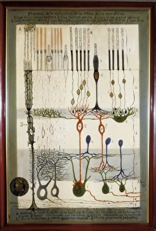
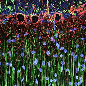
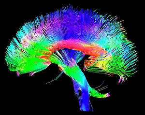
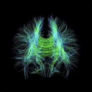
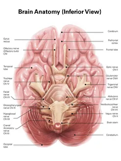
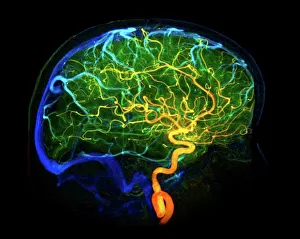
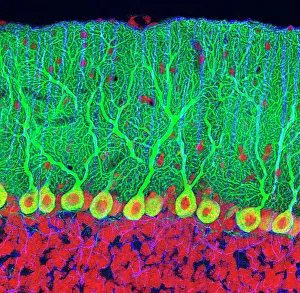
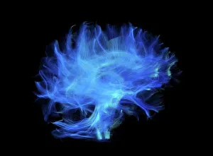
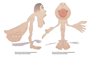
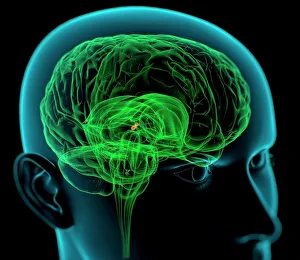
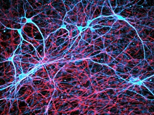
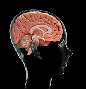
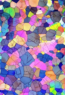
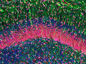
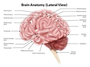
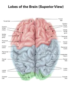
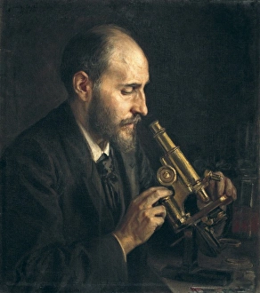
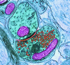
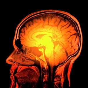
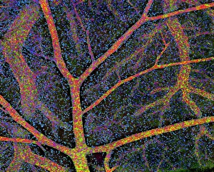
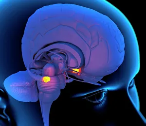
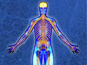
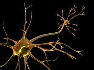
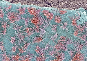
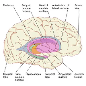
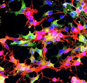
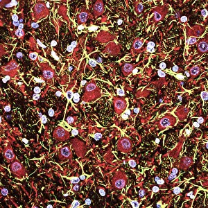
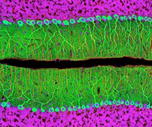
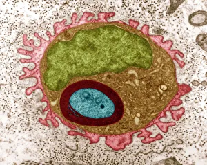
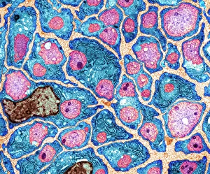
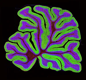
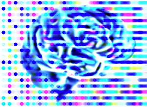
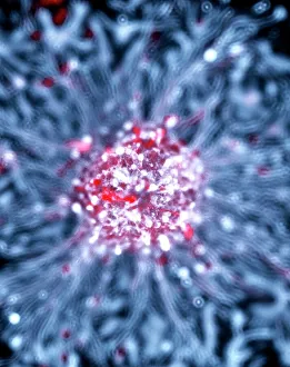
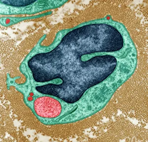
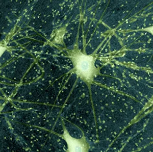
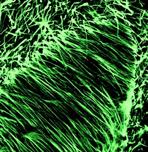
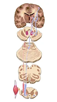
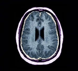
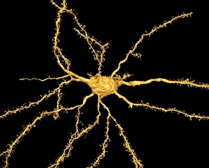
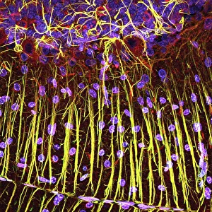
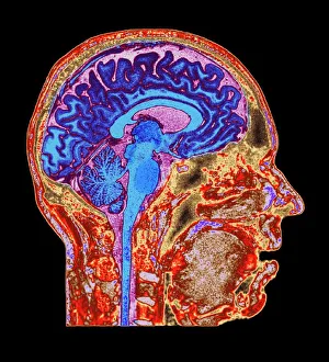
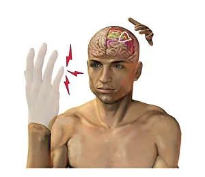
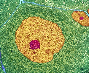
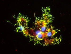
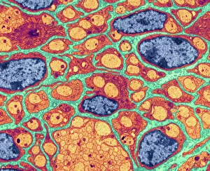
Neurology, the fascinating study of the nervous system, unravels the intricate workings of our brain and its various components. From the Motor homunculus model that depicts how different body parts are represented in our motor cortex to the Histological Diagram of a Mammalian Retina revealing the complexity of our visual processing, every aspect is awe-inspiring. The Cerebellum tissue, as seen through a light micrograph, showcases its unique structure responsible for coordinating movement and balance. Meanwhile, an inferior view of the Anatomy of human brain highlights its remarkable organization and interconnectedness. Through Brain fibres captured by DTI MRI scans like C017 / 7099 or C017 / 7035, we gain insights into neural pathways that facilitate communication between different regions. These pathways form a complex network crucial for transmitting information throughout our brain. Exploring Brain blood vessels using a 3D angiogram (C007 / 1981) reveals their vital role in supplying oxygen and nutrients to sustain neuronal function. Additionally, understanding Motor and sensory homunculi helps us comprehend how specific areas within our brains control different bodily functions. Delving deeper into neurology brings us face-to-face with Medulla oblongata artwork depicting this critical region involved in regulating essential autonomic functions such as breathing and heart rate. Furthermore, observing Nerve and glial cells under a light microscope illustrates their diverse roles in supporting neurons' health and functionality. Finally, examining Synapse nerve junctions through TEM unveils these microscopic structures where electrical signals are transmitted from one neuron to another—a fundamental process underlying all brain activity. In essence, neurology takes us on an incredible journey deep within ourselves—unveiling mysteries at both macroscopic levels like anatomical structures or microscopically exploring cellular intricacies, and is through this exploration that we gain profound knowledge about how our brains work—the very essence of who we are as individuals.

© Copyright Media Storehouse, All Rights Reserved 2003 - 2025
Facebook     Pinterest     100% Payment Secure     Reviews IO