Fraud Blocker Skip to main content

Nerve Fibre Collection (page 2)

"Nerve Fibres: Unveiling the Intricacies of Brain Connectivity" Delving into the depths of our complex neural network

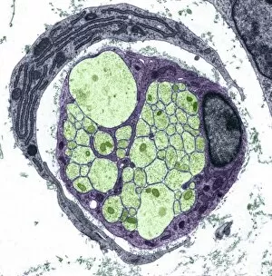
Background imageNerve Fibre Collection: Unmyelinated nerve, TEM

Unmyelinated nerve, TEM
Unmyelinated nerve. Transmission electron micrograph (TEM) of a section through axon (nerve fibre) bundles of unmyelinated nerves
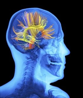
Background imageNerve Fibre Collection: Brain activity white matter fibres C016 / 6296

Brain activity white matter fibres C016 / 6296
White matter fibres overlaid a human head x-ray. Coloured 3D diffusion spectral imaging (DSI) scan of the bundles of white matter nerve fibres in the brain

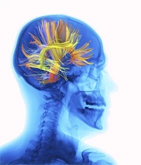
Background imageNerve Fibre Collection: Brain activity white matter fibres C016 / 6295

Brain activity white matter fibres C016 / 6295
White matter fibres overlaid a human head x-ray. Coloured 3D diffusion spectral imaging (DSI) scan of the bundles of white matter nerve fibres in the brain

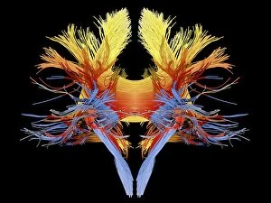
Background imageNerve Fibre Collection: White matter fibres of the human brain C018 / 6383

White matter fibres of the human brain C018 / 6383
White matter fibres. Computer enhanced 3D diffusion spectral imaging (DSI) scan of the bundles of white matter nerve fibres in the brain

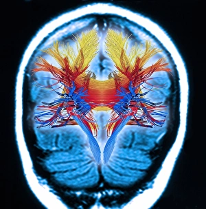
Background imageNerve Fibre Collection: White matter fibres, brain mri scan C018 / 6386

White matter fibres, brain mri scan C018 / 6386
White matter fibres overlaid a mri scan of the human head. Computer enhanced 3D diffusion spectral imaging (DSI) scan of the bundles of white matter nerve fibres in the brain

Background imageNerve Fibre Collection: White matter fibres of the human brain C018 / 6391

White matter fibres of the human brain C018 / 6391
White matter fibres. Computer enhanced 3D diffusion spectral imaging (DSI) scan of the bundles of white matter nerve fibres in the brain

Background imageNerve Fibre Collection: Corpus callosum, DTI MRI scan C017 / 7045

Corpus callosum, DTI MRI scan C017 / 7045
Corpus callosum. Coronal 3D diffusion tensor imaging (DTI) magnetic resonance imaging (MRI) scan of nerve pathways (blue) in and around the brains corpus callosum

Background imageNerve Fibre Collection: Glioblastoma brain tumour, DTI MRI scan C017 / 7052

Glioblastoma brain tumour, DTI MRI scan C017 / 7052
Glioblastoma brain tumour. 3D diffusion tensor imaging (DTI) magnetic resonance imaging (MRI) scan of nerve pathways (coloured) around a glioblastoma (upper right) in the brain

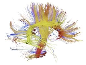
Background imageNerve Fibre Collection: White matter fibres of the human brain C014 / 5672

White matter fibres of the human brain C014 / 5672
White matter fibres. Coloured 3D diffusion spectral imaging (DSI) scan of the bundles of white matter nerve fibres in the brain

Background imageNerve Fibre Collection: White matter fibres of the human brain C014 / 5664

White matter fibres of the human brain C014 / 5664
White matter fibres. Coloured 3D diffusion spectral imaging (DSI) scan of the bundles of white matter nerve fibres in the brain

Background imageNerve Fibre Collection: White matter fibres of the human brain C014 / 5671

White matter fibres of the human brain C014 / 5671
White matter fibres. Coloured 3D diffusion spectral imaging (DSI) scan of the bundles of white matter nerve fibres in the brain

Background imageNerve Fibre Collection: White matter fibres of the human brain C014 / 5665

White matter fibres of the human brain C014 / 5665
White matter fibres. Coloured 3D diffusion spectral imaging (DSI) scan of the bundles of white matter nerve fibres in the brain

Background imageNerve Fibre Collection: White matter fibres of the human brain C014 / 5669

White matter fibres of the human brain C014 / 5669
White matter fibres. Coloured 3D diffusion spectral imaging (DSI) scan of the bundles of white matter nerve fibres in the brain

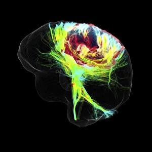
Background imageNerve Fibre Collection: White matter fibres and brain, artwork C015 / 1938

White matter fibres and brain, artwork C015 / 1938
White matter fibres overlaid a 3d model of the human brain in top view. Coloured 3D diffusion spectral imaging (DSI) scan of the bundles of white matter nerve fibres in the brain

Background imageNerve Fibre Collection: Myelinated nerve, TEM C016 / 5840

Myelinated nerve, TEM C016 / 5840
Myelinated nerve. Coloured transmission electron micrograph (TEM) of a section through a myelinated nerve fibre and Schwann cell

Background imageNerve Fibre Collection: Myelinated nerve, TEM C016 / 5839

Myelinated nerve, TEM C016 / 5839
Myelinated nerve. Coloured transmission electron micrograph (TEM) of a section through a myelinated nerve fibre and Schwann cell

Background imageNerve Fibre Collection: Myelinated nerve, TEM C016 / 5838

Myelinated nerve, TEM C016 / 5838
Myelinated nerve. Coloured transmission electron micrograph (TEM) of a section through a myelinated nerve fibre and Schwann cell

Background imageNerve Fibre Collection: Unmyelinated nerve, TEM C016 / 5805

Unmyelinated nerve, TEM C016 / 5805
Unmyelinated nerve. Transmission electron micrograph (TEM) of a section through axon (nerve fibre) bundles of unmyelinated nerves

Background imageNerve Fibre Collection: Unmyelinated nerve, TEM C016 / 5804

Unmyelinated nerve, TEM C016 / 5804
Unmyelinated nerve. Transmission electron micrograph (TEM) of a section through axon (nerve fibre) bundles of unmyelinated nerves

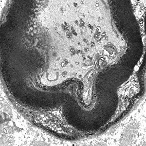
Background imageNerve Fibre Collection: Myelinated nerve, TEM C016 / 5448

Myelinated nerve, TEM C016 / 5448
Myelinated nerve. Transmission electron micrograph (TEM) of a section through a myelinated nerve fibre and Schwann cell. Myelin (black)

Background imageNerve Fibre Collection: Myelinated nerve, TEM C016 / 5370

Myelinated nerve, TEM C016 / 5370
Myelinated nerve. Transmission electron micrograph (TEM) of a section through a myelinated nerve fibre and Schwann cell (centre)

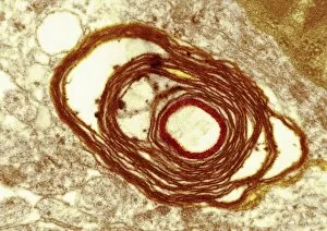
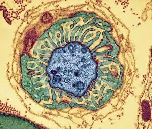
Background imageNerve Fibre Collection: Myelin surrounding a nerve axon, TEM

Myelin surrounding a nerve axon, TEM
Myelin surrounding a nerve axon, coloured transmission electron micrograph (TEM). The concentric round rings are the sheets of a Schwann cells myelin membrane (brown rings)

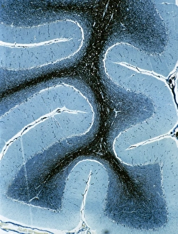
Background imageNerve Fibre Collection: LM of cortex and medulla of the cerebellum

LM of cortex and medulla of the cerebellum
Cerebellum. Light micrograph of a cross-section through the cortex (surface) and medulla (core) of a human cerebellum, part of the brain

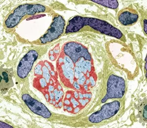
Background imageNerve Fibre Collection: Neurovascular bundle, TEM

Neurovascular bundle, TEM
Neurovascular bundle. Coloured transmission electron micrograph (TEM) of a cross section through a neurovascular bundle (NVB)

Background imageNerve Fibre Collection: Nerve fibre node, TEM

Nerve fibre node, TEM
Nerve fibre node. Coloured transmission electron micrograph (TEM) of a cross-section through a nerve fibre (axon) at a node of Ranvier

Background imageNerve Fibre Collection: Nerve fibres, TEM

Nerve fibres, TEM
Nerve fibres. Coloured transmission electron micrograph (TEM) of a section through non- myelinated nerve fibres (blue). The fibres are arranged into bundles surrounded by the cytoplasm of Schwann

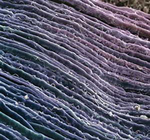
Background imageNerve Fibre Collection: Nerve fibres

Nerve fibres. Coloured scanning electron micrograph (SEM) of parallel myelinated nerve fibres in the spinal cord. Each fibre consists of a nerve cell axon, the output process of a nerve cell

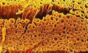
Background imageNerve Fibre Collection: Nerve fibre, SEM

Nerve fibre, SEM
Nerve fibres. Coloured scanning electron micrograph (SEM) of nerve fibres (brown). A group of nerves such as this is known as a fasciculus

Background imageNerve Fibre Collection: Coloured SEM of some nerve fibres

Coloured SEM of some nerve fibres
Nerve fibres. Coloured scanning electron micrograph (SEM) of some nerve fibres. A group of nerves such as this is known as a fasciculus

Background imageNerve Fibre Collection: False-colour SEM of a bundle of motor nerve fibres

False-colour SEM of a bundle of motor nerve fibres
Nerve fibres. False-colour scanning electron micrograph of a bundle of motor nerve fibres (blue). The bundle as a whole is surrounded by a cylindrical sheath of connective tissue

Background imageNerve Fibre Collection: Ilustration of an oligodendrocyte

Ilustration of an oligodendrocyte

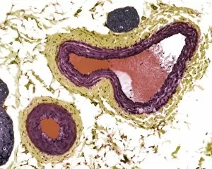
Background imageNerve Fibre Collection: Blood vessels, light micrograph

Blood vessels, light micrograph
Blood vessels. Light micrograph of a section through tissue showing an artery (bottom left) and a vein (top right). Two nerve fibres (dark blue) are also seen

Background imageNerve Fibre Collection: Nerve demyelination, TEM

Nerve demyelination, TEM
Nerve demyelination. Coloured transmission electron micrograph (TEM) of a section through a Schwann cell and a nerve fibre, showing the early collapse of its myelin sheath

Background imageNerve Fibre Collection: Multiple sclerosis

Multiple sclerosis. Illustration of the brain and spinal cord of a woman suffering from multiple sclerosis (MS). Also seen in the image is a magnified view of the spinal cord (upper right)

Background imageNerve Fibre Collection: Nerve fibres, light micrograph

Nerve fibres, light micrograph
Nerve fibres. Light micrograph of a transverse section through myelinated nerve fibres. The myelin sheaths are black, axons are blue. Magnification: x100 when printed at 10 centimetres wide

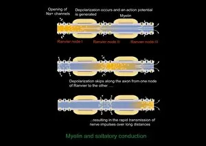
Background imageNerve Fibre Collection: Saltatory conduction of nerve impulse

Saltatory conduction of nerve impulse, computer artwork. Nerve impulses move along myelinated nerves by saltatory conduction. The myelin (orange, striped) does not cover the entire axon

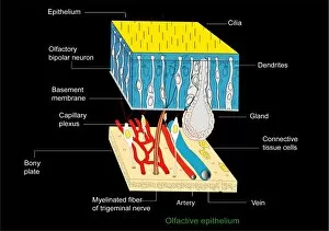
Background imageNerve Fibre Collection: Olfactory epithelium, artwork

Olfactory epithelium, artwork
Olfactory epithelium. Computer artwork showing the structure of the specialised layer of tissue that lines the inside of the nasal cavity and is involved in smell

Background imageNerve Fibre Collection: Nerve fibre, TEM

Nerve fibre, TEM
Nerve fibre. Coloured transmission electron micrograph (TEM) of a section through a non-myelinated peripheral nerve fibre

Background imageNerve Fibre Collection: Unmyelinated nerve bundle, TEM

Unmyelinated nerve bundle, TEM
Unmyelinated nerve bundle. Coloured transmission electron micrograph (TEM) of a section through a group of unmylinated nerve fibres

Background imageNerve Fibre Collection: Myelinated nerves, SEM

Myelinated nerves, SEM
Myelinated nerves. Coloured scanning electron micrograph (SEM) of a section through myelinated nerve fibres and Schwann cells


All Professionally Made to Order for Quick Shipping

"Nerve Fibres: Unveiling the Intricacies of Brain Connectivity" Delving into the depths of our complex neural network, nerve fibres serve as the intricate highways that facilitate communication within our brain. Through cutting-edge technology such as DTI MRI scans and immunofluorescent LM, scientists have begun unraveling the mysteries hidden within these delicate structures. The mesmerizing artwork depicting white matter fibres intertwined with the brain showcases their vital role in transmitting information across different regions. In C017 / 7099 and C017 / 7035 scans, we witness a captivating glimpse into this web-like connectivity, highlighting how these fibres enable seamless coordination between various cognitive processes. Immersing ourselves further into this microscopic realm, TEM images reveal regenerating nerve cells undergoing a remarkable transformation. Witnessing their resilience and ability to heal inspires awe at nature's incredible capacity for regeneration. Myelination emerges as another fascinating aspect captured by TEM imagery. The protective sheath surrounding nerve fibres ensures efficient transmission of electrical impulses, enhancing signal conduction speed and overall neural efficiency. As we explore diverse subjects like bee anatomy or artistic representations of white matter fibres in C014 / 5666 and C014 / 5668 artworks, we gain a deeper appreciation for both scientific inquiry and creative expression intertwining to shed light on these enigmatic structures. Through continued research on nerve fibre function and connectivity patterns, scientists strive to unlock new insights into neurological disorders such as Alzheimer's disease or multiple sclerosis. These investigations hold immense promise for developing targeted therapies aimed at preserving or restoring optimal brain function. Intricate yet resilient, nerve fibres stand as testaments to the complexity of human cognition. As science advances hand-in-hand with artistry in capturing their essence, humanity moves closer towards understanding one of its most profound creations – the human mind itself.